
Page 277 - Concise Pathology for Exam Preparation ( PDFDrive )
P. 277
262 SECTION II Diseases of Organ Systems
6. Infarct extension/expansion: New necrosis/weakening and disproportionate stretch-
ing, thinning or dilatation of infarct, leads to its extension/expansion
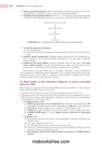
7. Embolism from mural thrombosis (Flowchart 11.5): Abnormal myocardial contractil-
ity leads to endocardial damage which in turn leads to mural thrombosis and embolism
Abnormal myocardial contractility
Endocardial damage
Mural thrombosis
Embolism
FLOWCHART 11.5. Pathogenesis of embolization from mural thrombosis.
8. Ventricular aneurysm formation:
(a) Late complication
(b) Associated with a large transmural anteroseptal infarct that converts into thin scar
tissue
9. Papillary muscle dysfunction: Postinfarct mitral regurgitation due to ischaemic in-
jury to papillary muscle and underlying myocardium; may later lead to papillary
muscle fibrosis.
10. Progressive late heart failure: Chronic ischaemic heart disease (also called isch-
aemic cardiomyopathy) is caused by postinfarction cardiac decompensation due to
exhaustion of compensatory hypertrophy of noninfarcted myocardium.
Complications occurring within first 72 h include cardiogenic shock, arrhythmias,
acute pulmonary oedema and cardiac tamponade. Late complications include cardiac an-
eurysm formation, Chronic IHD or ischaemic cardiomyopathy, congestive heart failure,
pulmonary hypertension and delayed pericarditis.
Q. Write briefly on the laboratory diagnosis of acute myocardial
infarction (MI).
Ans. A patient is diagnosed with myocardial infarction if two (probable) or three (definite)
of the following WHO criteria are met with:
• Clinical history of ischaemic type of chest pain lasting for more than 20 min.
• Changes in serial ECG tracings such as ST elevation/inverted T wave/appearance of Q wave.
• Rise in levels of serum cardiac biomarkers or enzymes, which leak out of the damaged
myocardium into the blood, such as:
1. Creatinine kinase (CK)
(a) Different isoenzymes of CK include MM (from skeletal muscle and heart), MB
(principally from myocardium, particularly MB2) and BB (from brain and lung).
(b) CK activity: Begins rising in 2–4 h, peaks in 24 h and falls in 72 h.
(c) CKMB: More specific/begins rising in 4–8 h, peaks in 18 h and falls in 48–72 h.
(d) CKMB2/CKMBI ratio .1.5 is a highly sensitive indicator of myocardial
injury.
2. Troponins (Tn)
(a) Troponins are proteins that regulate calcium mediated contraction of cardiac and
skeletal muscle.
(b) Two types, namely, TnI and TnT
(c) Not normally detectable in serum; elevated in acute MI
(d) Troponins of different origins can be distinguished by specific antibodies, which
can also be used for quantitative assays
(e) Most sensitive and specific cardiac markers; as sensitive as CKMB and more
specific
mebooksfree.com