
Page 321 - Concise Pathology for Exam Preparation ( PDFDrive )
P. 321
306 SECTION II Diseases of Organ Systems
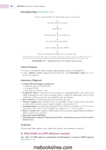
Pathophysiology (Flowchart 12.8)
Primary cytoskeletal defect of cell membrane (spectrin and ankyrin)
Decreased membrane stability
Membrane loss
Decreased surface area to volume ratio
Decreased cellular deformability*
Splenic trapping and stasis of RBCs
Extravascular haemolysis (phagocytosis and osmotic lysis)
*The discoid shape of the normal RBCs allows extreme degrees of deformation required to leave cords of Billroth and enter the
splenic sinusoids. Due to their spherical shape and limited deformability, spherocytes are sequestered in the splenic cords.
FLOWCHART 12.8. Pathophysiology of hereditary spherocytosis.
Clinical Features
• Presents in childhood with anaemia, splenomegaly, jaundice and gallstones
• Crises (aplastic crisis triggered by parvovirus B 19 and haemolytic crisis) are com-
monly encountered.
Laboratory Diagnosis
• General Haematological parameters:
• Reduced values of Hb and MCV
• Increased MCHC
• Increased reticulocyte count
• Peripheral smear: Anisocytosis with presence of microspherocytes and spherocytes
(dark appearing red cells with no central pallor). Unlike the spherocytes seen in other
conditions, spherocytes in HS are uniform in size and density
• Bone marrow: Erythroid hyperplasia (haemolytic picture)
• Osmotic fragility test: Determines the susceptibility of RBCs to haemolysis when they
are subjected to osmotic stress; osmotic fragility is increased in HS.
• Autohaemolysis: Spontaneous haemolysis when blood is incubated at 37°C for 48 h
(increased in HS; lysis of 10–15% RBCs as compared to , 4% in normal).
• Direct Coombs test: Negative (differentiates it from acquired spherocytosis of AIHA in
which Combs test is positive)
• Increased serum bilirubin
• Electrophoretic analysis of spectrin and other cytoskeleton protein levels (confir-
matory test)
Treatment
Splenectomy (after splenectomy, spherocytes persist but anaemia is corrected)
Q. Write briefly on G6PD deficiency anaemia.
Ans. Role of G6PD (glucose-6-phosphate dehydrogenase) enzyme in HMP pathway
(Flowchart 12.9):
mebooksfree.com