
Page 341 - Concise Pathology for Exam Preparation ( PDFDrive )
P. 341
326 SECTION II Diseases of Organ Systems
TABLE 12.15. Differences between acute lymphoblastic and acute myelogenous
leukaemia—cont’d
Acute lymphoblastic
Features leukaemia (ALL) Acute myelogenous leukaemia (AML)
Investigations
• Leukemic blasts Lymphoblasts (Fig. 12.5) Myeloblasts (Fig. 12.6)
• Size Smaller, 10–15 microns Larger, 12–20 microns
• N/C ratio High Low
• Chromatin Clumped Spongy, skein like
• Nucleoli < 2; indistinct 2–5; distinct
• Nuclear membrane Irregular, convoluted Regular
• Auer rods Not present Present in 10–20%
• TdT (terminal deoxynucleotidyl Often positive Negative
transferase)
Cytochemical staining
• Myeloperoxidase Negative Positive
• Sudan black B Negative Positive
• Chloroacetate esterase Negative Positive
• Periodic acid–Schiff (PAS) Positive (shows block pattern) Positive in < 25% of cells
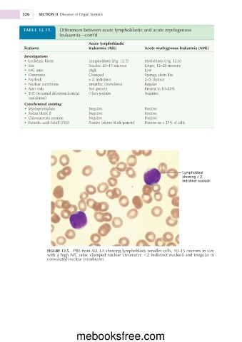
Lymphoblast
showing < 2
indistinct nucleoli
FIGURE 12.5. PBS from ALL L2 showing lymphoblasts (smaller cells, 10–15 microns in size
with a high N/C ratio; clumped nuclear chromatin; ,2 indistinct nucleoli and irregular to
convoluted nuclear membrane).
mebooksfree.com