
Page 371 - Concise Pathology for Exam Preparation ( PDFDrive )
P. 371
13
The Lung
AIRWAYS
Function: Exchange of gases between inspired air and blood
Histology: The entire respiratory tree is lined by pseudostratified tall columnar ciliated
epithelium admixed with mucous-secreting goblet cells in the cartilaginous airways.
Bronchial mucosa also has neuroendocrine cells that exhibit neurosecretory-type
granules, which contain serotonin, calcitonin and gastrin-releasing peptide.
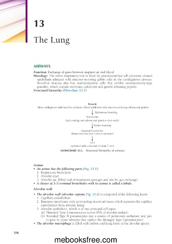
Structural hierarchy (Flowchart 13.1)
Bronchi
(have cartilaginous walls lined by columnar ciliated epithelium with mucous-producing submucosal glands)
Dichotomous branching
Bronchioles
(lack cartilage and submucosal glands in their walls)
Further branching
Terminal bronchioles
(bronchioles less than 2 mm in diameter)
Acini
(spherical with a diameter of about 7 mm)
FLOWCHART 13.1. Structural hierarchy of airways.
Acinus
• An acinus has the following parts (Fig. 13.1):
1. Respiratory bronchiole
2. Alveolar duct
3. Alveolar sac (blind end of respiratory passages and site for gas exchange)
• A cluster of 3–5 terminal bronchioles with its acinus is called a lobule.
Alveolar wall
• The alveolar wall (alveolar septum; Fig. 13.2) is composed of the following layers:
1. Capillary endothelium
2. Basement membrane with surrounding interstitial tissue which separates the capillary
endothelium from alveolar lining
3. Alveolar epithelium, which is of two principal cell types:
(a) Flattened Type I pneumocytes (cover 95% of alveolar surface)
(b) Rounded Type II pneumocytes (are a source of pulmonary surfactant and par-
ticipate in repair wherein they replace the damaged Type I pneumocytes)
• The alveolar macrophage is filled with carbon and lying loose in the alveolar spaces.
356
mebooksfree.com