
Page 374 - Concise Pathology for Exam Preparation ( PDFDrive )
P. 374
13 The Lung 359
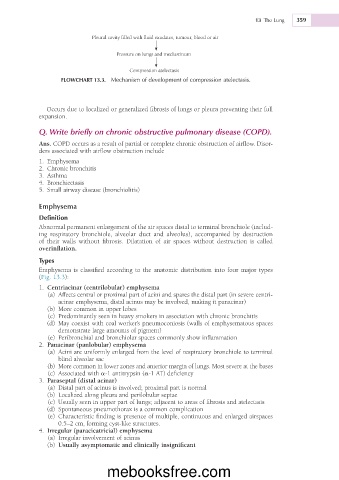
Pleural cavity filled with fluid exudates, tumour, blood or air
Pressure on lungs and mediastinum
Compression atelectasis
FLOWCHART 13.3. Mechanism of development of compression atelectasis.
Occurs due to localized or generalized fibrosis of lungs or pleura preventing their full
expansion.
Q. Write briefly on chronic obstructive pulmonary disease (COPD).
Ans. COPD occurs as a result of partial or complete chronic obstruction of airflow. Disor-
ders associated with airflow obstruction include
1. Emphysema
2. Chronic bronchitis
3. Asthma
4. Bronchiectasis
5. Small airway disease (bronchiolitis)
Emphysema
Definition
Abnormal permanent enlargement of the air spaces distal to terminal bronchiole (includ-
ing respiratory bronchiole, alveolar duct and alveolus), accompanied by destruction
of their walls without fibrosis. Dilatation of air spaces without destruction is called
overinflation.
Types
Emphysema is classified according to the anatomic distribution into four major types
(Fig. 13.3):
1. Centriacinar (centrilobular) emphysema
(a) Affects central or proximal part of acini and spares the distal part (in severe centri-
acinar emphysema, distal acinus may be involved, making it panacinar)
(b) More common in upper lobes
(c) Predominantly seen in heavy smokers in association with chronic bronchitis
(d) May coexist with coal worker’s pneumoconiosis (walls of emphysematous spaces
demonstrate large amounts of pigment)
(e) Peribronchial and bronchiolar spaces commonly show inflammation
2. Panacinar (panlobular) emphysema
(a) Acini are uniformly enlarged from the level of respiratory bronchiole to terminal
blind alveolar sac
(b) More common in lower zones and anterior margin of lungs. Most severe at the bases
(c) Associated with a-1 antitrypsin (a-1 AT) deficiency
3. Paraseptal (distal acinar)
(a) Distal part of acinus is involved; proximal part is normal
(b) Localized along pleura and perilobular septae
(c) Usually seen in upper part of lungs; adjacent to areas of fibrosis and atelectasis
(d) Spontaneous pneumothorax is a common complication
(e) Characteristic finding is presence of multiple, continuous and enlarged airspaces
0.5–2 cm, forming cyst-like structures.
4. Irregular (paracicatricial) emphysema
(a) Irregular involvement of acinus
(b) Usually asymptomatic and clinically insignificant
mebooksfree.com