
Page 373 - Concise Pathology for Exam Preparation ( PDFDrive )
P. 373
358 SECTION II Diseases of Organ Systems
• Burns
• Ionizing radiation
• Pulmonary embolization
• Inhalation of irritants
• Oxygen toxicity
• Smoke
• Gases and chemicals like ammonia, chlorine and nitrogen dioxide
• Chemical injury
• Heroin or methadone or barbiturate overdose
• Acetylsalicylic acid
• Thiazides
• Haematological conditions
• Multiple transfusions
• DIC
• Others
• Pancreatitis
• Uraemia
• Cardiopulmonary pass
• Hypersensitivity reactions
Gross Pathology
Heavy, red, boggy lungs, which ooze fluid on cutting
Microscopy
• Alveolar lining and pulmonary capillary endothelium are damaged.
• Alveolar walls are lined by a waxy hyaline membrane consisting of fibrin-rich oedema
fluid mixed with cytoplasmic and lipid remnants of necrotic epithelial cells.
• Type II pneumocytes proliferate to regenerate alveolar lining.
• Fibrin exudates organize and intra-alveolar fibrosis may ensue.
• Resolution is unusual; ARDS is commonly fatal.
X-ray
Shows diffuse bilateral infiltrates
Q. Define atelectasis. Enumerate and describe its various types.
Ans. Definition: Incomplete expansion of the lungs at birth (neonatal atelectasis) or collapse
of previously inflated lungs produces areas of relatively airless parenchyma.
Types:
1. Resorption atelectasis (Flowchart 13.2)
2. Compression atelectasis (Flowchart 13.3)
3. Contraction atelectasis:
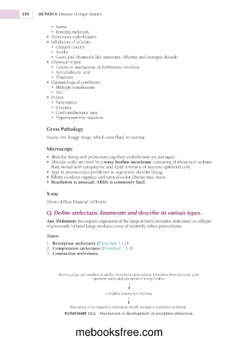
Mucous plugs and exudates in smaller bronchi (seen in asthma, bronchitis, bronchiectasis, post-
operative states) and aspiration of foreign bodies.
Complete obstruction of airway
Resorption of air trapped in dependent alveoli, leading to resorption atelectasis
FLOWCHART 13.2. Mechanism of development of resorption atelectasis.
mebooksfree.com