
Page 410 - Concise Pathology for Exam Preparation ( PDFDrive )
P. 410
14 The Oral Cavity and Gastrointestinal Tract 395
Clinical Features
Dysphagia/odynophagia, weight loss, iron deficiency, haemorrhage and sepsis from the
tumour, chest pain and vomiting.
Gross Morphology
60% polypoidal (fungating) lesions, 25% ulcerative and 15% diffuse infiltrating lesions.
Microscopy
• Majority are SCCs which involve the upper thoracic segment (half occurring in middle
third of oesophagus).
• May be superficial (carcinoma limited to mucosa and submucosa) or advanced (infiltrat-
ing into muscularis propria). Superficial lesions have a much better prognosis.
• Adenocarcinomas and undifferentiated carcinomas are less common.
• Adenocarcinomas usually arise in oesophageal mucous glands or Barrett’s oesophagus.
• Visceral metastasis to liver, lung and kidney is early and frequent.
• Overall prognosis is very poor.
STOMACH
Stomach is a saccular organ with a volume of about 1.5 L. It is divided into five anatomic
regions, each of which has different histology and functions. These are
1. Cardia: Where the contents of the oesophagus empty into the stomach
2. Fundus: Formed by the upper curvature of the organ
3. Body: Main central dome-shaped part
4. Pylorus: Lower part, which empties the contents of stomach into the small intestine
5. Pyloric sphincter: Stomach demarcated from the duodenum by this muscular sphincter
Layers of Stomach
• Mucosa: Consists of epithelium, lamina propria and a thin layer of smooth muscle
labelled muscularis mucosae
• Submucosa: Consists of fibrous connective tissue with the Meissner’s plexus
• Muscularis externa: Three layers of smooth muscle, namely:
• Inner oblique layer
• Middle circular layer
• Outer longitudinal layer
Auerbach’s plexus is found between the outer longitudinal layer and middle circular layer.
Normal gastric mucosa has two compartments:
1. Superficial foveolar, which is uniform throughout the stomach.
2. Deeper glandular compartment which has different types of cells found in the different
layers of these glands (Table 14.1).
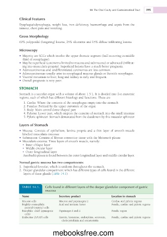
TABLE 14.1. Cells found in different layers of the deeper glandular component of gastric
mucosa
Name Secretory product Location in stomach
Mucous cells Mucous and pepsinogen II Cardiac and pyloric regions
Brightly eosinophilic Acid and intrinsic factor Fundic, cardiac and pyloric regions
parietal (oxyntic) cells
Basophilic chief (zymogenic Pepsinogen I and II Fundic region
cells)
Endocrine (APUD) cells Gastrin, histamine, endorphins, serotonin, Fundic, cardiac and pyloric regions
cholecystokinin and somatostatin
mebooksfree.com