
Page 435 - Concise Pathology for Exam Preparation ( PDFDrive )
P. 435
15
Diseases of the Hepatobiliary
System and Pancreas
LIVER
• Largest solid organ in the body (1200–1500 g)
• Divided into right and left lobes by the falciform ligament, the fissure of ligamentum
teres and the fissure of ligamentum venosum
• Surgical division into right and left hemilivers by the middle hepatic vein, which
lies between the inferior vena cava and the gallbladder, and passes through the porta
hepatis
• The right and left hemilivers are further subdivided into a total of 8 segments in accor-
dance with subdivisions of hepatic vasculature.
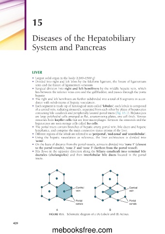
• Each segment is made up of histological units called ‘lobules’; each lobule is composed
of a central vein, radiating sinusoids, separated from each other by plates of hepatocytes
containing bile canaliculi and peripherally located portal tracts (Fig. 15.1). Hepatocytes
are large polyhedral cells arranged as flat, anastomosing plates, one cell thick. Venous
sinusoids have kupffer cells that are liver macrophages. Between the sinusoids and the
hepatocytes are seen storage cells called Ito cells.
• The portal tracts contain branches of hepatic artery, portal vein, bile ducts and hepatic
lymphatics, and comprise the main connective tissue stroma of the liver.
• Different regions of the lobule are referred to as ‘periportal’, ‘mid-zonal’ and ‘centrilobular’.
• Using the hepatic vasculature as reference, the liver architecture is divided into
‘acini’.
• On the basis of distance from the portal vessels, acinus is divided into ‘zone 1’ (closest
to the portal vessels), ‘zone 2’ and ‘zone 3’ (farthest from the portal vessel).
• Bile flows in the opposite direction along the biliary canaliculi into terminal bile
ductules (cholangioles) and then interlobular bile ducts located in the portal
tracts.
Central Central
vein vein
Portal Portal
canals canals
A B
FIGURE 15.1. Schematic diagram of a (A) Lobule and (B) Acinus.
420
420
mebooksfree.com