
Page 440 - Concise Pathology for Exam Preparation ( PDFDrive )
P. 440
15 Diseases of the Hepatobiliary System and Pancreas 425
3. Cholestatic (surgical) jaundice
(a) Cholestasis means failure of the bile flow and its cause may lie anywhere between
the hepatocyte and duodenum.
(b) Cholestasis can be due to small duct obstruction (intrahepatic cholestasis) or large
duct obstruction (extrahepatic cholestasis). Large bile duct obstruction is mainly
due to gallstones and malignancies of the head and neck of pancreas.
Indicators of cholestasis
- Hyperbilirubinaemia and bilirubinuria
- Elevated alkaline phosphatase activity
- Elevated gamma glutamyl transferase, 5 nucleotidase and leucine amino
peptidase
- Hypercholesterolaemia
- High serum bile salts (mainly cholate and chenodeoxycholate)
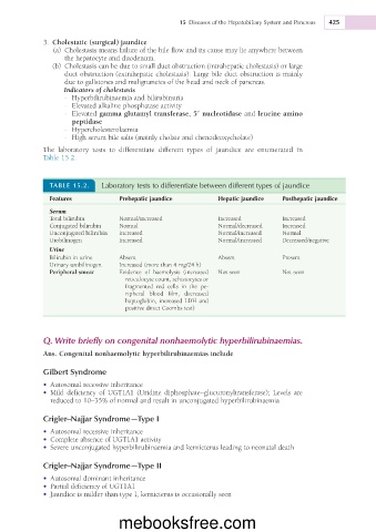
The laboratory tests to differentiate different types of jaundice are enumerated in
Table 15.2.
TABLE 15.2. Laboratory tests to differentiate between different types of jaundice
Features Prehepatic jaundice Hepatic jaundice Posthepatic jaundice
Serum
Total bilirubin Normal/increased Increased Increased
Conjugated bilirubin Normal Normal/decreased Increased
Unconjugated bilirubin Increased Normal/increased Normal
Urobilinogen Increased Normal/Increased Decreased/negative
Urine
Bilirubin in urine Absent Absent Present
Urinary urobilinogen Increased (more than 4 mg/24 h)
Peripheral smear Evidence of haemolysis (increased Not seen Not seen
reticulocyte count, schistocytes or
fragmented red cells in the pe-
ripheral blood film, decreased
haptoglobin, increased LDH and
positive direct Coombs test)
Q. Write briefly on congenital nonhaemolytic hyperbilirubinaemias.
Ans. Congenital nonhaemolytic hyperbilirubinaemias include
Gilbert Syndrome
• Autosomal recessive inheritance
• Mild deficiency of UGT1A1 (Uridine diphosphate–glucuronyltransferase); Levels are
reduced to 10–35% of normal and result in unconjugated hyperbilirubinaemia
Crigler–Najjar Syndrome—Type I
• Autosomal recessive inheritance
• Complete absence of UGT1A1 activity
• Severe unconjugated hyperbilirubinaemia and kernicterus leading to neonatal death
Crigler–Najjar Syndrome—Type II
• Autosomal dominant inheritance
• Partial deficiency of UGT1A1
• Jaundice is milder than type I, kernicterus is occasionally seen
mebooksfree.com