
Page 449 - Concise Pathology for Exam Preparation ( PDFDrive )
P. 449
434 SECTION II Diseases of Organ Systems
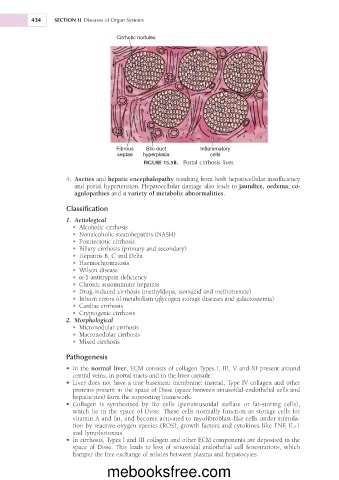
Cirrhotic nodules
Fibrous Bile duct Inflammatory
septae hyperplasia cells
FIGURE 15.5B. Portal cirrhosis liver.
4. Ascites and hepatic encephalopathy resulting from both hepatocellular insufficiency
and portal hypertension. Hepatocellular damage also leads to jaundice, oedema, co-
agulopathies and a variety of metabolic abnormalities.
Classification
1. Aetiological
• Alcoholic cirrhosis
• Nonalcoholic steatohepatitis (NASH)
• Postnecrotic cirrhosis
• Biliary cirrhosis (primary and secondary)
• Hepatitis B, C and Delta
• Haemochromatosis
• Wilson disease
• a-1 antitrypsin deficiency
• Chronic autoimmune hepatitis
• Drug-induced cirrhosis (methyldopa, isoniazid and methotrexate)
• Inborn errors of metabolism (glycogen storage diseases and galactosaemia)
• Cardiac cirrhosis
• Cryptogenic cirrhosis
2. Morphological
• Micronodular cirrhosis
• Macronodular cirrhosis
• Mixed cirrhosis
Pathogenesis
• In the normal liver, ECM consists of collagen Types I, III, V and XI present around
central veins, in portal tracts and in the liver capsule.
• Liver does not have a true basement membrane; instead, Type IV collagen and other
proteins present in the space of Disse (space between sinusoidal endothelial cells and
hepatocytes) form the supporting framework.
• Collagen is synthesized by Ito cells (perisinusoidal stellate or fat-storing cells),
which lie in the space of Disse. These cells normally function as storage cells for
vitamin A and fat, and become activated to myofibroblast-like cells under stimula-
tion by reactive oxygen species (ROS), growth factors and cytokines like TNF, IL-1
and lymphotoxins.
• In cirrhosis, Types I and III collagen and other ECM components are deposited in the
space of Disse. This leads to loss of sinusoidal endothelial cell fenestrations, which
hamper the free exchange of solutes between plasma and hepatocytes.
mebooksfree.com