
Page 450 - Concise Pathology for Exam Preparation ( PDFDrive )
P. 450
15 Diseases of the Hepatobiliary System and Pancreas 435
• Movement of proteins (albumin, clotting factors and lipoproteins) between plasma and
hepatocytes is markedly impaired, leading to functional changes in the liver.
Clinical Features
• Low-grade fever, weakness, fatigue, weight loss, anorexia, nausea, vomiting, upper
abdominal discomfort and abdominal distension due to ascites
• Menstrual irregularities like amenorrhea and irregular menses, hypogonadism, dimin-
ished body hair and gynaecomastia (due to impaired oestrogen metabolism and resulting
hyperestrogenaemia)
• Haemorrhagic tendencies like easy bruising, purpura, epistaxis, menorrhagia and gas-
trointestinal bleeding (decreased production of coagulation factors by the liver and
thrombocytopenia resulting from hypersplenism)
• Portal hypertension and its sequelae
Signs of Hepatocellular Failure
• Jaundice (due to abnormal bilirubin metabolism)
• Palmar erythema and spider naevi (due to localized vasodilatation)
• Parotid enlargement (attributed to fatty infiltration since liver’s ability to break down
body fat is reduced in cirrhosis)
• Ascites (due to portal hypertension; and low levels of albumin in the blood)
• Hepatic encephalopathy and flapping tremors (associated with increased blood ammo-
nia levels)
• Progressive renal dysfunction (due to decreased renal perfusion attributed to systemic
vasodilatation)
Q. Outline the aetiopathogenesis and clinical features of portal
hypertension.
Ans. Portal hypertension is defined as a clinical condition in which there is prolonged
elevation of portal venous pressure due to increased resistance to portal blood flow.
Causes
• Prehepatic: Portal vein thrombosis and fibrosis of bile ducts (schistosomiasis)
• Intrahepatic: Cirrhosis, schistosomiasis, massive fatty change, sarcoidosis and miliary
tuberculosis
• Posthepatic: Obstruction of hepatic vein by thrombosis (Budd–Chiari syndrome) or tumours
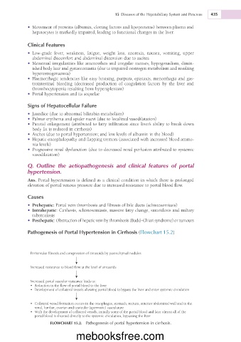
Pathogenesis of Portal Hypertension in Cirrhosis (Flowchart 15.2)
Perivenular fibrosis and compression of sinusoids by parenchymal nodules
Increased resistance to blood flow at the level of sinusoids
Increased portal vascular resistance leads to:
• Reduction in the flow of portal blood to the liver
• Development of collateral vessels allowing portal blood to bypass the liver and enter systemic circulation
• Collateral vessel formation occurs in the oesophagus, stomach, rectum, anterior abdominal wall and in the
renal, lumbar, ovarian and testicular (spermatic) vasculature
• With the development of collateral vessels, initially some of the portal blood and later almost all of the
portal blood is shunted directly to the systemic circulation, bypassing the liver
FLOWCHART 15.2. Pathogenesis of portal hypertension in cirrhosis.
mebooksfree.com