
Page 481 - Concise Pathology for Exam Preparation ( PDFDrive )
P. 481
466 SECTION II Diseases of Organ Systems
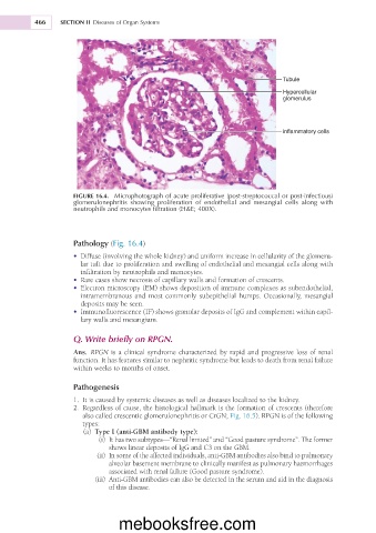
Tubule
Hypercellular
glomerulus
inflammatory cells
FIGURE 16.4. Microphotograph of acute proliferative (post-streptococcal or post-infectious)
glomerulonephritis showing proliferation of endothelial and mesangial cells along with
neutrophils and monocytes filtration (H&E; 400X).
Pathology (Fig. 16.4)
• Diffuse (involving the whole kidney) and uniform increase in cellularity of the glomeru-
lar tuft due to proliferation and swelling of endothelial and mesangial cells along with
infiltration by neutrophils and monocytes.
• Rare cases show necrosis of capillary walls and formation of crescents.
• Electron microscopy (EM) shows deposition of immune complexes as subendothelial,
intramembranous and most commonly subepithelial humps. Occasionally, mesangial
deposits may be seen.
• Immunofluorescence (IF) shows granular deposits of IgG and complement within capil-
lary walls and mesangium.
Q. Write briefly on RPGN.
Ans. RPGN is a clinical syndrome characterized by rapid and progressive loss of renal
function. It has features similar to nephritic syndrome but leads to death from renal failure
within weeks to months of onset.
Pathogenesis
1. It is caused by systemic diseases as well as diseases localized to the kidney.
2. Regardless of cause, the histological hallmark is the formation of crescents (therefore
also called crescentic glomerulonephritis or CrGN; Fig. 16.5). RPGN is of the following
types:
(a) Type I (anti-GBM antibody type):
(i) It has two subtypes—“Renal limited” and “Good pasture syndrome”. The former
shows linear deposits of IgG and C3 on the GBM.
(ii) In some of the affected individuals, anti-GBM antibodies also bind to pulmonary
alveolar basement membrane to clinically manifest as pulmonary haemorrhages
associated with renal failure (Good pasture syndrome).
(iii) Anti-GBM antibodies can also be detected in the serum and aid in the diagnosis
of this disease.
mebooksfree.com