
Page 486 - Concise Pathology for Exam Preparation ( PDFDrive )
P. 486
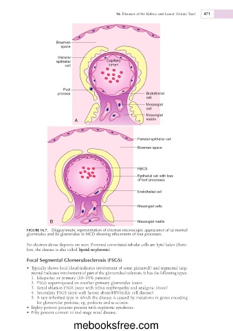
16  Diseases of the Kidney and Lower Urinary Tract  471


                          Bowman
                            space

                           Visceral
                          epithelial             Capillary
                              cell                lumen


                             Post
                           process                                Bndothelial
                                                                  cell
                                                                  Mesangial
                                                                  cell
                                                                  Mesongial
                                  A                               matrix


                                                               Parietal epithelial cell
                                                               Bowman space


                                                               RBCS
                                                               Epithelial cell with loss
                                                               of toot processes

                                                               Endothelial cell


                                                               Mesangial cells


                       B                                       Mesangial matrix
             FIGURE 16.7.  Diagrammatic representation of electron microscopic appearance of (a) normal
             glomerulus and (b) glomerulus in MCD showing effacement of foot processes.


             No electron-dense deposits are seen. Proximal convoluted tubular cells are lipid laden (there-
             fore, the disease is also called lipoid nephrosis).

             Focal Segmental Glomerulosclerosis (FSGS)
             •  Typically shows focal (focal indicates involvement of some glomeruli) and segmental (seg-
               mental indicates involvement of part of the glomerulus) sclerosis. It has the following types:
                1.  Idiopathic or primary (10–35% patients)
                2.  FSGS superimposed on another primary glomerular lesion
                3.  Renal ablation FSGS (seen with reflux nephropathy and analgesic abuse)
                4.  Secondary FSGS (seen with heroin abuse/HIV/sickle cell disease)
                5.  A rare inherited type in which the disease is caused by mutations in genes encoding
                  for glomerular proteins, eg, podocin and a-actinin.
             •  Eighty percent patients present with nephrotic syndrome.
             •  Fifty percent convert to end-stage renal disease.


                                  mebooksfree.com