
Page 482 - Concise Pathology for Exam Preparation ( PDFDrive )
P. 482
16 Diseases of the Kidney and Lower Urinary Tract 467
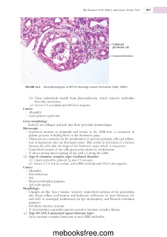
Collapsed
glomerular tuft
Crescent formation
FIGURE 16.5. Microphotograph of RPGN showing cresent formation (H&E; 400X).
(iv) These individuals benefit from plasmapheresis, which removes antibodies
from the circulation.
(v) Serum C3 is normal and ANCA is negative.
Causes:
• Idiopathic
• Good pasture syndrome
Gross morphology:
Kidneys are enlarged and pale and show petechial haemorrhages.
Microscopy:
• Segmental necrosis in glomeruli and breaks in the GBM lead to exudation of
plasma proteins including fibrin in the Bowman’s space.
• Fibrin acts as a stimulus for the proliferation of parietal epithelial cells and infiltra-
tion of monocytes into the Bowman’s space. This results in formation of crescents
because the cells take the shape of the Bowman’s space which is crescentic).
• Uninvolved portion of the cells glomerulus shows no proliferation.
• IF shows strong linear staining of IgG and C3 along the GBM.
(b) Type II (immune complex type) mediated disorder:
(i) Characterized by granular Ig and C3 deposits
(ii) Serum C3 is low to normal, anti-GBM antibody and ANCA are negative.
Causes:
• Idiopathic
• Post-infectious
• SLE
• Henoch–Schönlein purpura
• IgA nephropathy
Morphology:
• Changes are like Type I disease, however, uninvolved portions of the glomerulus
also shows diffuse proliferation and leukocyte infiltration (in post-infectious GN
and SLE) or mesangial proliferation (in IgA nephropathy and Henoch–Schönlein
purpura).
• EM shows discrete deposits.
• IF demonstrates a granular pattern typical of immune complex disease.
(c) Type III ANCA-associated (pauci-immune type)
Lacks immune complex formation or anti-GBM antibodies.
mebooksfree.com