
Page 499 - Concise Pathology for Exam Preparation ( PDFDrive )
P. 499
484 SECTION II Diseases of Organ Systems
TABLE 16.12. Clinicopathological features of the most common types of RCC—cont’d
Collecting duct
Chromophobe (Bellini duct) Xp11 transloca-
Features Clear-cell RCC Papillary RCC RCC carcinoma tion carcinoma
Gross • Solitary, unilat- Multifocal, bilat- • Tan brown Seen in medullary -
eral, bright yel- eral, less yellow • Excellent region
low to grey- due to lower prognosis
white with lipid content,
prominent cys- papillae may be
tic change and seen, haemor-
haemorrhage rhagic and cys-
• Aggressive; may tic areas present
infiltrate into
surrounding
substance, col-
lecting system,
calyces, ureters
and renal vein
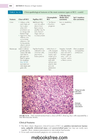
Microscopy Solid to tubular Papillae lined by, Solid sheets of Irregular channels Clear cytoplasm
growth pattern, cuboidal to low cells arranged lined by malig- with papillary
round cells with columnar cells; around blood nant cells with a architecture
clear (due to psammoma vessels, indi- hobnail appear-
glycogen and bodies present vidual cell is ance; cells en-
lipid) or granu- eosinophilic meshed within
lar cytoplasm with well- a fibrotic stroma
(Fig. 16.10); defined cyto-
may show nu- plasmic mar-
clear atypia and gins and
giant cells perinuclear
halo
Polygonal cells
with clear
cytoplasm
Delicate
branching
vasculature
FIGURE 16.10. H&E-stained section from a clear-cell RCC showing clear cells separated by a
fine fibrovascular stroma.
Clinical Features
• The three classic diagnostic clinical features of RCC are painless intermittent haema-
turia, palpable abdominal mass and costovertebral pain but they are rarely seen
together. Most common presentation is intermittent haematuria.
• Fever and constitutional symptoms are commonly seen.
mebooksfree.com