
Page 498 - Concise Pathology for Exam Preparation ( PDFDrive )
P. 498
16 Diseases of the Kidney and Lower Urinary Tract 483
• Also called ‘hypernephroma’ due to resemblance to clear cells of adrenal cortex and
gross yellow colour
• Arises from tubular epithelium (renal adenocarcinomas)
Epidemiology
• Predisposing factors: Smoking, obesity, hypertension, unopposed oestrogen therapy,
exposure to asbestos, cadmium, petroleum products and heavy metals and acquired
cystic disease in patients with long-standing dialysis.
• Majority of cases of RCC are sporadic; about 5% are inherited and associated with:
1. von Hippel–Lindau (VHL) syndrome: Predisposition to a large number of neo-
plasms, mainly haemangioblastomas of cerebellum and retina, multiple bilateral
renal cysts, pheochromocytomas and multicentric bilateral renal cell carcinomas.
2. Hereditary leiomyomatosis and renal cell cancer syndrome: Autosomal dominant
inheritance; mutation in Fumarate Hydratase (FH) gene; associated with uterine and
cutaneous leiomyomas and an aggressive variety of papillary RCC.
3. Hereditary papillary RCC: Autosomal dominant inheritance; multiple cytogenetic
abnormalities; mutation in MET proto-oncogene; associated with multiple bilateral
papillary RCCs.
4. Birt–Hogg–Dube (BHD) syndrome: Autosomal dominant inheritance; mutation in BHD
gene (expresses folliculin); associated with skin appendageal tumours of hair follicular
origin, pulmonary cysts and renal tumours.
Gross Morphology
• Globular, encapsulated, 3–5 cm, soft, lobulated with a variegated appearance
(grey-white to yellow with necrosis, haemorrhage and cyst formation); invades or grows
into pelvis.
• Polar in distribution; the upper pole is more commonly involved than the lower pole.
• Renal vascular invasion is common.
• Usually sharply defined; however, small satellite nodules are often found in the
surrounding substance.
• Enlarges n bulges into pelvis and calyces n fungates through walls of collecting system
n ureters.
• Penetrates through capsule n invades perinephric fat and adrenals.
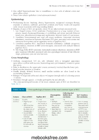
The clinicopathological features of the most common types of RCC are described in
Table 16.12.
TABLE 16.12. Clinicopathological features of the most common types of RCC
Collecting duct
Chromophobe (Bellini duct) Xp11 transloca-
Features Clear-cell RCC Papillary RCC RCC carcinoma tion carcinoma
Incidence 70–80% 10–15% 5–8% 1%
Genetics • Majority spo- • Culprit (tyro- • Multiple • Several chro- • Seen in young
radic. sine kinase re- chromosomal mosomal ab- patients and is
• 98% show loss ceptor for the loss and hy- normalities associated with
of material on hepatocytic podiploidy seen but no overexpression
the short arm of growth factor) • Arise from in- definite pattern of TFE3 tran-
chromosome 3 is on chromo- tercalated cells recognized scription factor
• Second allele some 7q31 lining collect- due to translo-
lost by somatic • Duplication of ing ducts cations of TFE3
mutation chromosome 7 gene located at
• Loss of both increases gene Xp11.2 with a
copies of VHL dosage of MET number of other
gene gives rise oncogene lead- genes.
to clear-cell car- ing to abnormal
cinoma growth of distal
tubular cells
Continued
mebooksfree.com