
Page 497 - Concise Pathology for Exam Preparation ( PDFDrive )
P. 497
482 SECTION II Diseases of Organ Systems
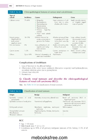
TABLE 16.10. Clinicopathological features of various renal calculi/stones
Type of
calculi Incidence Causes Pathogenesis Gross
Calcium stones 75–80% • Idiopathic Super saturation of cal- Small, smooth contour,
• Hypercalciuria cium ions in urine, or irregular jagged
• Hypercalcaemia alkaline pH of urine mass of spicules
• Hyperoxaluria
• Hyperuricosuria
• Primary hyperthy-
roidism
• Distal renal tubular
acidosis
Struvite stones 10–15% Urinary infection by Alkaline urinary pH due Large, solitary, branch-
[MgNH 4 (PO) 3 ] urease-containing or- to production of am- ing structure formed
triple stone/ ganisms like Proteus monia from urea (by due to progressive
stag-horn stone urease) accretion of salts
Uric acid stones 6% Gout, dehydration, id- Acidic urine and g solu- Smooth, yellow to
iopathic and malig- bility of uric acid brownish, hard and
nant tumours multiple
Cystine stones 1–2% Hereditary Cystine precipitates in Small, smooth yellow,
acidic urine multiple and round
Others Up to 10% Inherited abnormality of Xanthinuria
amino acid metabolism
Complications of Urolithiasis
1. Loss of function in the affected kidney
2. Obstruction of the ureter (acute unilateral obstructive uropathy) and hydronephrosis;
secondary infection gives rise to pyonephrosis
3. Urinary tract infection
4. Haematuria
Q. Classify renal tumours and describe the clinicopathological
features of renal cell carcinoma (RCC).
Ans. See Table 16.11 for classification of renal tumours.
TABLE 16.11. Classification of renal tumours
Origin Benign Malignant
Epithelial tumours of renal Adenoma, oncocytoma, adrenal rests Renal cell carcinoma (RCC or
parenchyma hypernephroma)
Epithelial tumours of renal pelvis Transitional cell papilloma Transitional cell carcinoma (TCC),
squamous cell carcinoma, adeno-
carcinoma of renal pelvis
Embryonal tumours Mesoblastic nephroma, multicystic Wilms tumour
nephroma
Nonepithelial tumours Angiomyolipoma, fibroma, leiomyoma Sarcoma
Miscellaneous Reninoma –
Metastatic tumours – –
RCC
• Age: . 60 years
• Male:female ratio 5 2:1 to 3:1
• Constitutes up to 90% of all primary malignant tumours of the kidney, 2–3% of all
cancers.
mebooksfree.com