
Page 534 - Concise Pathology for Exam Preparation ( PDFDrive )
P. 534
18 Female Genital System 519
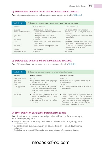
Q. Differentiate between serous and mucinous ovarian tumours.
Ans. Differences between serous and mucinous ovarian tumours are listed in Table 18.2.
TABLE 18.2. Differences between serous and mucinous ovarian tumours
Features Serous tumours Mucinous tumours
Frequency Most common ovarian tumour Less common than serous tumours
Incidence of malignancy Account for 60% of all malignant ovarian Account for 10% of malignant ovarian
tumours tumours
Age affected • Benign lesions: 30–40 years, Middle age; rare before puberty and after
• Malignant lesions: 45–65 years menopause
Bilateralism Common Less/rare
Gross Unilocular/few cysts filled with clear se- Multilocular tumours filled with sticky
rous fluid gelatinous fluid rich in glycoproteins
Cell lining Tall columnar ciliated epithelial cells Tall columnar cells resembling endocer-
vical or intestinal epithelium
Papillae Very common Less common
Psammoma bodies Common Not found
Q. Differentiate between mature and immature teratoma.
Ans. Differences between mature and immature teratoma are listed in Table 18.3.
TABLE 18.3. Differences between mature and immature teratoma
Features Mature teratoma Immature teratoma
Component tissue Mature Immature
Age affected Young women (reproductive age group) Adolescents and young adults (before age 20)
Bilateralism Bilateral in 10–15% cases Mostly unilateral
Type Mostly cystic (dermoid cyst) Usually solid
Gross appearance Unilocular cyst lined by the epidermis. Predominantly solid with areas of necrosis and
Cyst may have areas of calcification, haemorrhage
teeth, matted hair and sebaceous ma-
terial
Microscopy • Cyst wall lined by mature stratified • Immature structures differentiating towards
squamous epithelium with appenda- cartilage, glands, muscles, bones, neuroepi-
geal structures. thelium, etc., seen. Tissue resembles fetal or
• No immature elements/neuroepithe- embryonic tissue rather than adult tissue.
lium seen • Proportion of immature neuroepithelium in
tumour determines the prognosis
Q. Write briefly on gestational trophoblastic disease.
Ans. Gestational trophoblastic disease usually develops within uterus, but may develop at
any site of ectopic pregnancy.
• Ranges in behaviour from benign hydatidiform mole (H. mole) to highly aggressive
choriocarcinoma.
• All secrete human chorionic gonadotropin (HCG), which can be detected in the serum
and urine.
• The fall or rise in titres of HCG can be used as an indicator of response to therapy.
mebooksfree.com