
Page 530 - Concise Pathology for Exam Preparation ( PDFDrive )
P. 530
18 Female Genital System 515
- Psammoma bodies in 32% cases (thought to be associated with a better
survival)
- Five-year survival ,20%
(b) Mucinous tumours
• Common in the reproductive age group
• Eighty percent benign, 10% of low malignant potential and 10% are frankly
malignant.
• Five percent benign and 20% malignant tumours are bilateral.
• Larger and more multilocular than their serous counterparts.
• Papillary formations and psammoma bodies less common than their serous
counterparts.
• Lined by tall columnar epithelium with a basal nucleus and abundant cytoplasmic
mucin (cells similar to endocervical mucosa).
• Multiloculated cysts filled with sticky or gelatinous mucinous material.
• Glistening, smooth and papery thin wall.
• Solid areas or papillary projections on inner wall of the cyst suggestive of malignant
change.
(i) Benign mucinous cystadenoma
Gross morphology:
Multilocular thin-walled cysts containing sticky gelatinous material.
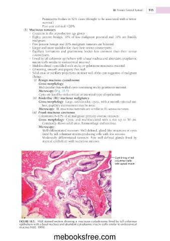
Microscopy (Fig. 18.5):
Cysts are lined by endocervical or intestinal type of epithelium
(ii) Borderline (BL) mucinous malignancy
Gross morphology: Large, multilocular, cystic, with a smooth external sur-
face; papillary excrescences may be seen.
Microscopy: BL mucinous tumours are similar to BL serous tumours.
(iii) Frank mucinous carcinoma
Constitutes 6–10% of all malignant primary ovarian tumours.
Gross morphology: Cystic and multiloculated with a size up to 50 cm.
Commonly shows solid areas, haemorrhage and necrosis.
Microscopy:
- Well-differentiated tumours: Well-defined, gland-like structures or cysts
lined by tall columnar mucin-producing cells with few mitoses.
- Moderately differentiated tumours: Few well-defined glands lined by
atypical epithelium with numerous mitoses.
Cyst lining of tall
columnar cells
with apical mucin
FIGURE 18.5. H&E-stained section showing a mucinous cystadenoma lined by tall columnar
epithelium with a basal nucleus and abundant cytoplasmic mucin (cells similar to endocervical
mucosa H&E; 100X).
mebooksfree.com