
Page 535 - Concise Pathology for Exam Preparation ( PDFDrive )
P. 535
520 SECTION II Diseases of Organ Systems
H. Mole
• Traditionally discovered during 12–14 weeks of pregnancy.
• Uterine enlargement is more than what is anticipated for that period of gestation.
• Manifests with vaginal bleeding and passage of grape-like tissue mass.
• Elevation of HCG (particularly the beta subunit) in blood and urine and absence of fetal
parts or fetal heart sound on sonography is diagnostic.
• It is of two types, namely, complete and partial mole.
Gross Morphology
• Uterine cavity/ectopic site is filled with delicate, friable masses of thin-walled, translucent,
cystic and grape-like structures.
• Amniotic sac is very small and collapsed.
• No fetal parts in complete mole; may be seen in partial mole.
Microscopic Examination
• Complete mole
• All villi show hydropic swelling and complete loss of vascularity.
• The central substance of the villi is loose, myxomatous and oedematous, covered by
a layer of chorionic epithelium (cytotrophoblast and syncytiotrophoblast).
• Villi show circumferential proliferation of epithelium to produce sheets and masses of
the same.
• Partial mole
• Villous oedema restricted to some villi.
• Trophoblastic proliferation is mild and focal.
• Villi have a characteristic irregular scalloped margin.
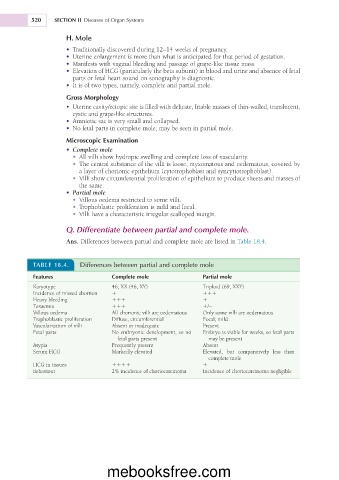
Q. Differentiate between partial and complete mole.
Ans. Differences between partial and complete mole are listed in Table 18.4.
TABLE 18.4. Differences between partial and complete mole
Features Complete mole Partial mole
Karyotype 46, XX (46, XY) Triploid (69, XXY)
Incidence of missed abortion 1 111
Heavy bleeding 111 1
Toxaemia 111 1/–
Villous oedema All chorionic villi are oedematous Only some villi are oedematous
Trophoblastic proliferation Diffuse; circumferential Focal; mild
Vascularization of villi Absent or inadequate Present
Fetal parts No embryonic development, so no Embryo is viable for weeks, so fetal parts
fetal parts present may be present
Atypia Frequently present Absent
Serum HCG Markedly elevated Elevated, but comparatively less than
complete mole
HCG in tissues 1111 1
Behaviour 2% incidence of choriocarcinoma Incidence of choriocarcinoma negligible
mebooksfree.com