
Page 565 - Concise Pathology for Exam Preparation ( PDFDrive )
P. 565
550 SECTION II Diseases of Organ Systems
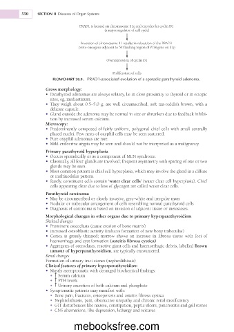
PRAD1 is located on chromosome 11q and encodes for cyclin D1
(a major regulator of cell cycle)
Inversion of chromosome 11 results in relocation of the PRAD1
proto-oncogene adjacent to 5¢ flanking region of PTH gene on 11p
Overexpression of cyclin D1
Proliferation of cells
FLOWCHART 20.9. PRAD1-associated evolution of a sporadic parathyroid adenoma.
Gross morphology:
• Parathyroid adenomas are always solitary, lie in close proximity to thyroid or in ectopic
sites, eg, mediastinum.
• They weigh about 0.5–5.0 g, are well circumscribed, soft tan-reddish brown, with a
delicate capsule.
• Gland outside the adenoma may be normal in size or shrunken due to feedback inhibi-
tion by increased serum calcium.
Microscopy:
• Predominantly composed of fairly uniform, polygonal chief cells with small centrally
placed nuclei. Few nests of oxyphil cells may be seen scattered.
• Pure oxyphil adenomas are rare.
• Mild endocrine atypia may be seen and should not be interpreted as a malignancy.
Primary parathyroid hyperplasia
• Occurs sporadically or as a component of MEN syndrome.
• Classically, all four glands are involved; frequent asymmetry with sparing of one or two
glands may be seen.
• Most common pattern is chief cell hyperplasia, which may involve the gland in a diffuse
or multinodular pattern.
• Rarely, constituent cells contain ‘water clear cells’ (water clear cell hyperplasia). Chief
cells appearing clear due to loss of glycogen are called water clear cells.
Parathyroid carcinoma
• May be circumscribed or clearly invasive, grey-white and irregular mass
• Nodular or trabecular arrangement of cells resembling normal parathyroid cells
• Diagnosis of carcinoma is based on invasion of adjacent tissue or metastases.
Morphological changes in other organs due to primary hyperparathyroidism
Skeletal changes
• Prominent osteoclasts (cause erosion of bone matrix)
• Increased osteoblastic activity (induces formation of new bony trabeculae)
• Cortex is grossly thinned; marrow shows an increase in fibrous tissue with foci of
haemorrhage and cyst formation (osteitis fibrosa cystica)
• Aggregates of osteoclasts, reactive giant cells and haemorrhagic debris, labelled Brown
tumour of hyperparathyroidism, are typically encountered.
Renal changes
Formation of urinary tract stones (nephrolithiasis)
Clinical features of primary hyperparathyroidism:
• Mostly asymptomatic with deranged biochemical findings:
h
• Serum calcium
h
• PTH levels
h
• Urinary excretion of both calcium and phosphate
• Symptomatic patients may manifest with:
• Bone pain, fractures, osteoporosis and osteitis fibrosa cystica
• Nephrolithiasis, pain, obstructive uropathy and chronic renal insufficiency
• GIT disturbances like nausea, constipation, peptic ulcers, pancreatitis and gall stones
• CNS alternations, like depression, lethargy and seizures
mebooksfree.com