
Page 561 - Concise Pathology for Exam Preparation ( PDFDrive )
P. 561
546 SECTION II Diseases of Organ Systems
translocation between chromosomes 10 and 17 formation of RET/PTC
fusion gene or NTRK1 fusion gene activation of MAP kinase pathway.
• Mutations in signal transduction genes (RAS mutations and mutations in BRAF
oncogene).
(c) Medullary carcinoma
• Sporadic in 80% cases; remainder occur in a setting of MEN IIA or IIB or as familial
tumours not associated with MEN syndrome.
• Familial tumours occurring in MEN Type II are associated with germline muta-
tions in RET protooncogene which leads to constitutive activation of tyrosine
kinase receptor and cellular proliferation.
2. Environmental factors
• Association with ionizing radiation
• Pre-existing thyroid pathology, eg, nodular goitre, adenomas and Hashimoto thyroiditis.
Papillary Thyroid Carcinoma
Clinical features
• Most common thyroid malignancy
• Peak incidence between 20 and 40 years; may be seen at any age
• Presents as a solitary (cold) nodule
• In most cases, primary thyroid nodule is asymptomatic and cervical lymph node metastasis
is the first manifestation.
• Primary thyroid nodule may sometimes manifest with hoarseness, dysphagia, cough
and dyspnoea.
Predisposing factors
• Previous exposure to ionizing radiation
• Increased incidence of PTC is observed in Gardner syndrome (familial adenomatous
polyposis coli) and Cowden disease (familial goitre and skin haematomas)
Gross morphology:
• Solitary or multifocal; often cystic
• May be well circumscribed/encapsulated or ill-defined/infiltrative
• On cut surface, papillary areas are easily identified and appear granular. Areas of fibrosis
may be seen
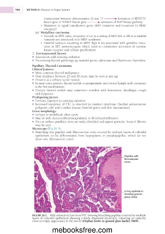
Microscopy (Fig 20.3):
• Branching true papillae with fibrovascular cores covered by multiple layers of cuboidal
epithelium (to be differentiated from hyperplastic or pseudopapillae, which do not
show true fibrovascular cores).
Papillae with
fibrovascular
cores
Lining epithelium
showing ground
glass nuclei
FIGURE 20.3. H&E-stained section from PTC showing branching papillae covered by multiple
layers of cuboidal epithelium showing a finely dispersed chromatin, imparting an optically
clear or empty appearance to the nuclei (Orphan Annie or ground glass nuclei; 100X).
mebooksfree.com