
Page 564 - Concise Pathology for Exam Preparation ( PDFDrive )
P. 564
20 Endocrinology 549
• Yellow-brown, ovoid, encapsulated, measuring 35–40 mg
• Composed of:
• Chief cells (secrete PTH or parathormone)
• Oxyphil cells (appear at the onset of puberty, but have no known function)
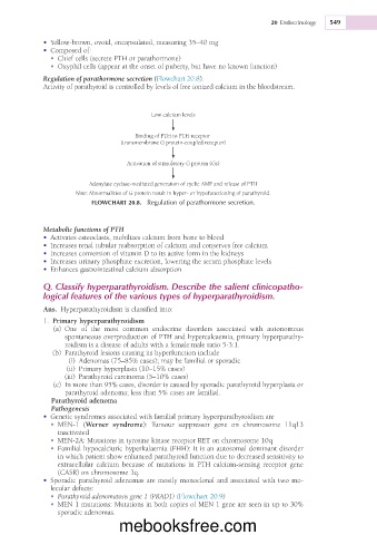
Regulation of parathormone secretion (Flowchart 20.8).
Activity of parathyroid is controlled by levels of free ionized calcium in the bloodstream.
Low calcium levels
Binding of PTH to PTH receptor
(transmembrane G protein-coupled receptor)
Activation of stimulatory G protein (Gs)
Adenylate cyclase-mediated generation of cyclic AMP and release of PTH
Note: Abnormalities of G protein result in hyper- or hypofunctioning of parathyroid.
FLOWCHART 20.8. Regulation of parathormone secretion.
Metabolic functions of PTH
• Activates osteoclasts, mobilizes calcium from bone to blood
• Increases renal tubular reabsorption of calcium and conserves free calcium
• Increases conversion of vitamin D to its active form in the kidneys
• Increases urinary phosphate excretion, lowering the serum phosphate levels
• Enhances gastrointestinal calcium absorption
Q. Classify hyperparathyroidism. Describe the salient clinicopatho-
logical features of the various types of hyperparathyroidism.
Ans. Hyperparathyroidism is classified into:
1. Primary hyperparathyroidism
(a) One of the most common endocrine disorders associated with autonomous
spontaneous overproduction of PTH and hypercalcaemia, primary hyperparathy-
roidism is a disease of adults with a female:male ratio 5-3:1.
(b) Parathyroid lesions causing its hyperfunction include
(i) Adenomas (75–85% cases); may be familial or sporadic
(ii) Primary hyperplasia (10–15% cases)
(iii) Parathyroid carcinoma (5–10% cases)
(c) In more than 95% cases, disorder is caused by sporadic parathyroid hyperplasia or
parathyroid adenoma; less than 5% cases are familial.
Parathyroid adenoma
Pathogenesis
• Genetic syndromes associated with familial primary hyperparathyroidism are
• MEN-1 (Werner syndrome): Tumour suppressor gene on chromosome 11q13
inactivated
• MEN-2A: Mutations in tyrosine kinase receptor RET on chromosome 10q
• Familial hypocalciuric hyperkalaemia (FHH): It is an autosomal dominant disorder
in which patient show enhanced parathyroid function due to decreased sensitivity to
extracellular calcium because of mutations in PTH calcium-sensing receptor gene
(CASR) on chromosome 3q.
• Sporadic parathyroid adenomas are mostly monoclonal and associated with two mo-
lecular defects:
• Parathyroid adenomatosis gene 1 (PRAD1) (Flowchart 20.9)
• MEN 1 mutations: Mutations in both copies of MEN 1 gene are seen in up to 30%
sporadic adenomas.
mebooksfree.com