
Page 606 - Concise Pathology for Exam Preparation ( PDFDrive )
P. 606
21 Musculoskeletal System 591
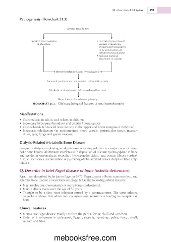
Pathogenesis (Flowchart 21.3)
Chronic renal failure
Impaired renal excretion • Decreased conversion of
of phosphate vitamin D metabolite
25-hydroxycholecalciferol
to its active form 1,25-
dihydroxycholecalciferol
• Reduced intestinal
absorption of calcium
Hyperphosphataemia and hypocalcaemia
Increased parathormone and resultant osteoclastic activity
Metabolic acidosis (result of decreased renal function)
Major lesions of renal osteodystrophy
FLOWCHART 21.3. Clinicopathological features of renal osteodystrophy
Manifestations
• Osteomalacia in adults and rickets in children
• Secondary hyperparathyroidism and osteitis fibrosa cystica
• Osteosclerosis (enhanced bone density in the upper and lower margins of vertebrae)
• Metastatic calcification (in medium-sized blood vessels, periarticular tissue, myocar-
dium, eyes, lungs and gastric mucosa)
Dialysis-Related Metabolic Bone Disease
Long-term dialysis employing an aluminium-containing solution is a major cause of meta-
bolic bone lesions (aluminium interferes with deposition of calcium hydroxyapatite in bone
and results in osteomalacia, secondary hyperparathyroidism and osteitis fibrosa cystica).
Also, in such cases, accumulation of b 2 -microglobulin amyloid causes dialysis-related amy-
loidosis.
Q. Describe in brief Paget disease of bone (osteitis deformans).
Ans. First described by Sir James Paget in 1877; Paget disease of bone is an osteolytic and
sclerotic bone disease of uncertain aetiology. It has the following salient features:
• May involve one (monostotic) or more bones (polyostotic).
• Mainly affects males over the age of 50 years.
• Thought to be a slow virus infection caused by a paramyxovirus. The virus infested
osteoclasts release IL-6 which induces osteoclastic recruitment leading to resorption of
bone.
Clinical Features
• Monostotic Paget disease mainly involves the pelvis, femur, skull and vertebrae.
• Order of involvement in polyostotic Paget disease is: vertebrae, pelvis, femur, skull,
sacrum and tibia.
mebooksfree.com