
Page 608 - Concise Pathology for Exam Preparation ( PDFDrive )
P. 608
21 Musculoskeletal System 593
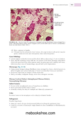
Benign
fibroblastic
tissue
Curvilinear bony
trabeculae of
woven bone
FIGURE 21.18. Section from FD composed of benign-looking fibroblastic tissue arranged in a
loose, whorled pattern within which irregular and curved trabeculae of woven (nonlamellar)
bone are laid down (H&E; 1003).
(d) More common in females
(e) Characterized by polyostotic bone lesions, skin pigmentation (café-au-lait macular
spots), sexual precocity and infrequently other endocrinopathies.
Gross Pathology
• Lesions appear as sharply demarcated localized defects measuring 2–5 cm in diameter.
• Thin, smooth overlying cortex with the cut section of the lesion showing replacement
of normal cancellous bone of the marrow cavity by gritty, grey-pink, rubbery soft tissue,
which may have areas of haemorrhage, myxoid and cystic degeneration.
Microscopy (Fig. 21.18)
• Characteristic benign-looking fibroblastic tissue arranged in a loose, whorled pattern in
which irregular and curved trabeculae of woven (nonlamellar) bones are laid down.
• Numerous osteoclasts in relation to bony trabeculae.
• Rarely, secondary malignant change, most often osteogenic sarcoma.
Fibrous Cortical Defect (Metaphyseal Fibrous Defect,
Nonossifying Fibroma)
Salient Features
• Occurs in the metaphyseal cortex of long bones in children
• Most commonly involves tibia or femur
• Generally solitary, but may be multiple and bilaterally symmetrical
X-Ray
Eccentric lesion in the metaphysis with a sharply delimited border
Pathogenesis
Possible hypothesis:
• Arises as a result of some developmental defects involving the epiphyseal plate
• Could be a tumour of histiocytic origin (based on close resemblance to fibrohistiocytic
tumours)
mebooksfree.com