


Page 67 - Concise Pathology for Exam Preparation ( PDFDrive )
P. 67
52    SECTION I  General Pathology


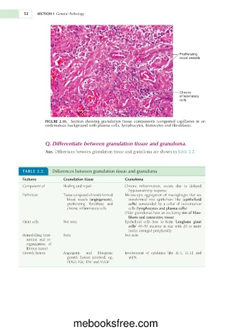
                                                                                  Proliferating
                                                                                  blood vessels


                                                                                  Chronic
                                                                                  inflammatory
                                                                                  cells


                     FIGURE  2.10.  Section  showing  granulation  tissue  components  (congested  capillaries  in  an
                     oedematous background with plasma cells, lymphocytes, histiocytes and fibroblasts).


                     Q. Differentiate between granulation tissue and granuloma.

                     Ans. Differences between granulation tissue and granuloma are shown in Table 2.2.


           TABLE 2.2.    Differences between granulation tissue and granuloma

           Features           Granulation tissue         Granuloma
           Component of       Healing and repair         Chronic  inflammation;  occurs  due  to  delayed
                                                           hypersensitivity response
           Definition         Tissue composed of newly formed   Microscopic aggregation of macrophages that are
                               blood  vessels  (angiogenesis),   transformed  into  epithelium  like  (epithelioid
                               proliferating  fibroblasts  and   cells)  surrounded  by  a  collar  of  mononuclear
                               chronic inflammatory cells  cells (lymphocytes and plasma cells)
                                                         Older granulomas have an enclosing rim of fibro-
                                                           blasts and connective tissue
           Giant cells        Not seen                   Epithelioid  cells  fuse  to  form  ‘Langhans  giant
                                                           cells’  40–50  microns  in  size  with  20  or  more
                                                           nuclei arranged peripherally
           Remodelling (mat-  Seen                       Not seen
             uration  and  re-
             organization  of
             fibrous tissue)
           Growth factors     Angiogenic   and   fibrogenic   Involvement  of  cytokines  like  IL-1,  IL-12  and
                               growth  factors  involved,  eg,   gIFN
                               PDGF, FGF, TNF and VEGF


                                  mebooksfree.com

