


Page 64 - Concise Pathology for Exam Preparation ( PDFDrive )
P. 64
2  Acute and Chronic Inflammation  49


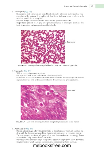
               3.  Eosinophil (Fig. 2.6)
                •  Recruitment and extravasation from blood driven by adhesion molecules like neu-
                  trophils  and  by  eotaxin  (chemokine  derived  from  leukocytes  and  epithelial  cells
                  which is specific for eosinophils)
                •  Involved in IgE-mediated immune reactions and parasitic infections
                •  Major basic protein is a highly toxic protein contained in eosinophil granules. It is
                  toxic to parasites and mammalian epithelial cells.


                                                                    Eosinophils


                   FIGURE 2.6.  Eosinophil showing a bilobed nucleus and coarse red granules.


               4.  Mast cells (Fig. 2.7)
                •  Widely present in connective tissue.
                •  Participate in both acute and chronic inflammatory cells.
                •  Express on their surface the receptor that binds to the Fc portion of IgE antibody to
                  degranulate mast cells and release mediators (histamines and prostaglandins).


                                                                     Mast cells


                 FIGURE 2.7.  Mast cells showing abundant basophilic granules and round nuclei.


               5.  Plasma cells (Fig. 2.8)
                •  Plasma cells are large cells with amphophilic to basophilic cytoplasm, an eccentric nu-
                  cleus with the chromatin arranged in a characteristic cart-wheel or clock-face pattern.
                •  Their cytoplasm contains a pale perinuclear zone that on electron microscopy shows
                  an extensive Golgi apparatus and centrioles.
                •  Plasma cells have abundant rough endoplasmic reticulum coupled with a well-developed
                  Golgi apparatus, which together are responsible for immunoglobulin secretion.
                                  mebooksfree.com

