


Page 65 - Concise Pathology for Exam Preparation ( PDFDrive )
P. 65
50    SECTION I  General Pathology


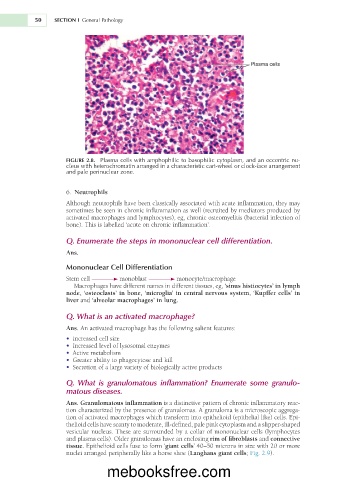
                                                                              Plasma cells


                     FIGURE 2.8.  Plasma cells with amphophilic to basophilic cytoplasm, and an eccentric nu-
                     cleus with heterochromatin arranged in a characteristic cart-wheel or clock-face arrangement
                     and pale perinuclear zone.


                       6.  Neutrophils
                     Although neutrophils have been classically associated with acute inflammation, they may
                     sometimes be seen in chronic inflammation as well (recruited by mediators produced by
                     activated macrophages and lymphocytes), eg, chronic osteomyelitis (bacterial infection of
                     bone). This is labelled ‘acute on chronic inflammation’.

                     Q. Enumerate the steps in mononuclear cell differentiation.

                     Ans.
                     Mononuclear Cell Differentiation

                     Stem cell        monoblast         monocyte/macrophage
                        Macrophages have different names in different tissues, eg, ‘sinus histiocytes’ in lymph
                     node, ‘osteoclasts’ in bone, ‘microglia’ in central nervous system, ‘Kupffer cells’ in
                     liver and ‘alveolar macrophages’ in lung.

                     Q. What is an activated macrophage?
                     Ans. An activated macrophage has the following salient features:
                     •  Increased cell size
                     •  Increased level of lysosomal enzymes
                     •  Active metabolism
                     •  Greater ability to phagocytose and kill
                     •  Secretion of a large variety of biologically active products

                     Q.  What  is  granulomatous  inflammation?  Enumerate  some  granulo-
                     matous diseases.

                     Ans. Granulomatous inflammation is a distinctive pattern of chronic inflammatory reac-
                     tion characterized by the presence of granulomas. A granuloma is a microscopic aggrega-
                     tion of activated macrophages which transform into epithelioid (epithelial like) cells. Epi-
                     thelioid cells have scanty to moderate, ill-defined, pale pink cytoplasm and a slipper-shaped
                     vesicular nucleus. These are surrounded by a collar of mononuclear cells (lymphocytes
                     and plasma cells). Older granulomas have an enclosing rim of fibroblasts and connective
                     tissue. Epithelioid cells fuse to form ‘giant cells’ 40–50 microns in size with 20 or more
                     nuclei arranged peripherally like a horse shoe (Langhans giant cells; Fig. 2.9).


                                  mebooksfree.com

