


Page 66 - Concise Pathology for Exam Preparation ( PDFDrive )
P. 66
2  Acute and Chronic Inflammation  51


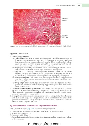
                                                                          Caseous
                                                                          necrosis


                                                                          Langhans
                                                                          giant cell


             FIGURE 2.9.  A caseating epithelioid cell granuloma with Langhans giant cells (H&E; 100x).


             Types of Granulomas

               1.  Infectious granulomas
                 (a)  Tuberculosis (prototype of granulomatous disease): Caused by Mycobacterium tu-
                   berculosis, tuberculosis is associated with the formation of caseating granulomas
                   (granulomas showing presence of central granular debris with loss of all cellular
                   detail and higher positivity for acid fast bacilli) or noncaseating granulomas (ab-
                   sence of caseation and low positivity for acid fast bacilli).
                 (b)  Leprosy: It is caused by Mycobacterium leprae. Noncaseating granulomas are typi-
                   cally seen with or without acid fast lepra bacilli in the macrophages.
                 (c)  Syphilis:  It  is  caused  by  Treponema  pallidum.  Gumma  formation  is  the  disease
                   hallmark. Gumma is histopathologically characterized by a central necrotic area
                   without loss of cellular outline; plasma cell infiltrate with a wall of histiocytes.
                  (d)  Cat scratch disease: It is caused by a Gram-negative bacillus. It typically shows
                   rounded or stellate granulomas containing central granular debris and large num-
                   ber of neutrophils.
                 (e)  Deep fungal infections: Fungal granulomas are caused by organisms like histo-
                   plasma  and  blastomyces  and  are  typically  suppurative  (granulomas  with  neutro-
                   philic inflammation).
               2.  Noninfectious or immune granulomas: Granulomas form in response to persistent
                presence of nondegradable or particulate material, which incites an immune response.
                These are usually noncaseating epithelioid cell granulomas. Examples includes arcoid-
                osis and hypersensitivity pneumonitis.
               3.  Foreign body granulomas are formed as a response to foreign bodies like talc, suture
                and  intravenous  drugs.  The  foreign  material  can  be  identified  in  the  centre  of  the
                granuloma or within the foreign body giant cells which have a haphazard distribution
                of nuclei unlike Langhans giant cell.
             Q. Enumerate the components of granulation tissue.

             Ans. Granulation tissue (Fig. 2.10) has the following components:
               1.  Newly formed blood vessels (endothelial proliferation or neoangiogenesis)
               2.  Chronic inflammatory cells
               3.  Proliferating fibroblasts
              4.  Extracellular matrix which in comparison to ordinary extracellular matrix is more cellular
                and more vascular


                                  mebooksfree.com

