


Page 89 - Concise Pathology for Exam Preparation ( PDFDrive )
P. 89
74    SECTION I    General Pathology


                                                                                 Congested
                                                                                 capillaries
                                                                                 in alveolar
                                                                                 wall


                                                                                 Heart failure
                                                                                 cells


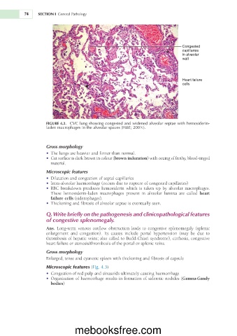
                     FIGURE 4.2.    CVC lung showing congested and widened alveolar septae with hemosiderin-
                     laden macrophages in the alveolar spaces (H&E; 2003).


                     Gross morphology
                        •	�The   lungs   are   heavier   and   firmer   than   normal.
                                                                            o
                                                                             f

                                 s
                        •	�Cut  surface    dark  brown    colour  (brown induration)  with  oozing    frothy,  blood-tinged
                                            n
                                            i
                                i
                       material.
                     Microscopic features
                        •	�Dilatation   and   congestion     septal   capillaries
                                             of
                                                                 of
                        •	�Intra-alveolar   haemorrhage   (occurs   due     rupture     congested   capillaries)
                                                        to


                                                              is


                        •	�RBC  breakdown  produces  hemosiderin  which      taken  up  by  alveolar  macrophages.


                       These    hemosiderin-laden  macrophages  present  in  alveolar  lumina  are  called  heart


                       failure cells   (siderophages).
                                           of
                                                         is
                        •	�Thickening   and   fibrosis     alveolar   septae     eventually   seen.
                     Q.   Write briefly on the pathogenesis and clinicopathological features
                     of congestive splenomegaly.


                                                                to

                     Ans.    Long-term  venous  outflow  obstruction  leads      congestive  splenomegaly  (splenic


                     enlargement    and  congestion).  Its  causes  include  portal  hypertension  (may  be  due  to


                                                      to
                               of
                     thrombosis     hepatic   veins;   also   called     Budd-Chiari   syndrome),   cirrhosis,   congestive
                                or
                                                  of
                                                             or
                     heart   failure     stenosis/thrombosis     the   portal     splenic   veins.
                     Gross morphology
                     Enlarged,   tense   and   cyanotic   spleen   with   thickening   and   fibrosis     capsule
                                                                          of
                     Microscopic features   (Fig. 4.3)
                                 of
                        •	�Congestion     red   pulp   and   sinusoids   ultimately   causing   haemorrhage
                                   f
                                                                 f
                                                     i
                                                      n
                                                                o

                        •	�Organization     haemorrhage  results    formation     siderotic  nodules (Gamna-Gandy


                                  o
                       bodies)
                                  mebooksfree.com

