


Page 91 - Concise Pathology for Exam Preparation ( PDFDrive )
P. 91
76 SECTION I General Pathology
of
or
perfused, undergoes centrilobular necrosis with loss drop out hepatocytes. Severe
long-standing congestion may lead grossly evident hepatic fibrosis (cardiac cirrhosis).
to
Q. Define and describe types of haemorrhage. Write in detail on
haemostasis.
of
to
is
Ans. Extravasation red cells due vessel rupture called haemorrhage.
may
is
or
it
• � It be external (external haemorrhage) enclosed within the tissue (when
a
called haematoma).
• �Haematoma may insignificant (bruise) large enough cause fatality, eg, a large
be
or
to
an
retroperitoneal haematoma internal (visceral) haematoma.
or
or
• �Minute 1–2 mm haemorrhages into the skin, mucous membranes serosal surfaces are
as
denoted petechiae (typically seen with locally increased intravascular pressure, low
platelet counts and defective platelet function).
as
• �Larger .3 mm haemorrhages are called purpura (associated with the same disorders
to
or
petechiae and also occur secondary trauma, vasculitis increased vascular fragility).
• �Still larger .1–2 cm subcutaneous haematomas or bruises are called ecchymoses
(generally seen after trauma).
• �Accumulation blood body cavities may called haemothorax, haemopericardium,
b
o
e
n
i
f
haemoperitoneum and haemarthrosis (joints) depending on the cavity involved.
Haemostasis
is
Haemostasis the mechanism which maintains the integrity the circulatory system after
of
of
vascular damage. Normal haemostatic mechanism the body has three components:
Vascular component: This involves reflex spasm the injured vessel (vasoconstriction),
o
a
1
f
.
which minimizes the blood loss.
.
Platelet component: Platelets are anucleate discoid structures derived from marrow
2
o
f
megakaryocytes. The cytoplasm platelets contains three major types storage granules:
f
o
a
(i) Alpha granules containing variety proteins like fibrinogen and von Willebrand factor
o
f
(ii) Dense granules containing serotonin, ADP and calcium
(iii) Lysosomal granules containing acid hydrolases
t
o
o
t
Following vessel constriction, platelets adhere the vessel wall and also aggregate
a
form platelet plug which seals off the vascular breach and arrests haemorrhage (primary
i
s
f
haemostasis). This followed activation the coagulation cascade and fibrin deposition
o
y
b
(secondary haemostasis).
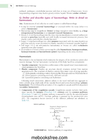
3. cascade includes three path-
Components of the coagulation cascade: Coagulation
ways, namely, the intrinsic (Flowchart 4.4), extrinsic and the common pathways
in
is
(Flowcharts 4.5 and 4.6). Intrinsic pathway assessed vitro by the activated partial
is
thromboplastin time (aPTT). Extrinsic pathway assessed by the prothrombin time
(PT). The coagulation factors involved in the different pathways are tabulated in
Table 4.4.
Intrinsic pathway
Negatively charged particles
Contact activation
HMW kininogen
XII XIIa
XI XIa
Thrombin
IX IXa
VIIIa
VIII
Ca ++
Platelets
X Xa
FLOWCHART 4.4. Intrinsic pathway of coagulation.
mebooksfree.com

