


Page 94 - Concise Pathology for Exam Preparation ( PDFDrive )
P. 94
4  Haemodynamic Disorders, Thrombosis and Shock   79

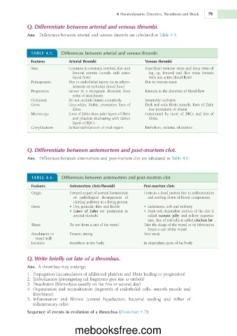
             Q.   Differentiate between arterial and venous thrombi.

                                                                     in
             Ans.     Differences   between   arterial   and   venous   thrombi   are   tabulated     Table   4.5.


               TABLE   4.5.   Differences between arterial and venous thrombi
               Features        Arterial thrombi	�                Venous thrombi
               Sites           Common  in      cerebral,   iliac   and     Superficial   varicose   veins   and   deep   veins  of
                                         coronary,


                                 femoral    arteries  (vessels  with  active     leg,    eg,  femoral  and  iliac  veins  (vessels


                                 blood   flow)                     with   less   active   blood   flow)
                                     endothelial
                                                                       venous
               Pathogenesis    Due  to       injury   (as  in    Due  to    stasis
                                                      athero-
                                          turbulent
                                 sclerosis  or    blood   flow)
                                                                         the

                                                                                    blood
               Progression     Grows    in  a  retrograde  direction  from     Extends  in    direction  of    flow


                                 point  of
                                        attachment
               Occlusion       Do   not   occlude   lumen   completely   Invariably   occlusive


               Gross           Grey-white,   friable,  prominent  lines  of   Dark   red  with  fibrin  strands;  lines  of  Zahn


                                 Zahn                              less   prominent  or
                                                                                absent
                                      Zahn
               Microscopy      Lines  of    show   paler   layers  of      Constituted    by  more  of  RBCs  and  less  of


                                                        fibrin


                                 and   platelets  alternating  with  darker     fibrin
                                        RBCs
                                 layers  of
                                                vital
               Complications   Ischaemia/infarction  of    organs   Embolism,   oedema,   ulceration
             Q.   Differentiate between antemortem and post-mortem clot.
             Ans.     Differences   between   antemortem   and   post-mortem   clot   are   tabulated     Table   4.6.
                                                                          in
               TABLE   4.6.   Differences between antemortem and post-mortem clot
               Features         Antemortem clots/thrombi        Post-mortem clots
                                                                                      sedimentation
                                        part  of
                                                                          a  dead
               Origin           Formed  as      normal   haemostasis     Form  in    person   due  to

                                                                                 blood

                                 or    pathological  derangement  of     and   settling   down  of    components
                                                  living
                                 clotting   pathway  in  a    person
               Gross               •  Dry,   granular,   firm   and   friable     •  Gelatinous,   soft   and   rubbery
                                   •  Lines  of  Zahn  are  prominent  in       •  Dark   red,   dependent   portion  of    clot  is


                                                                                         the

                                 arterial   thrombi               called    currant  jelly  and  yellow  superna-


                                                                           red
                                                                                   called
                                                                  tant,   free  of    cells  is    chicken fat
                                                                              the
                                                                                        bifurcation
                                           cast  of
               Shape            Do   not   form  a      the   vessel   Take   the   shape  of    vessel  or  its
                                                                  forms      a  cast  of    vessel
                                                                             the
               Attachment  to   Present;   strong               Very  weak

                 vessel   wall
               Location         Anywhere  in    body            In   dependent   parts  of    body
                                                                                the
                                          the
             Q.   Write briefly on fate of a thrombus.
             Ans.     thrombus   may   undergo:
                  A
                                      of
                                                                       to
             1.
                 Propagation   (accumulation     additional   platelets   and   fibrin   leading     progression)
             2.            (propagating   tail   fragments   give   rise     emboli)
                 Embolization
                                                        to
                                                   or
             3.           (fibrinolysis   usually   on   the   first     second   day)
                 Dissolution


             4.                                        endothelial  cells,  smooth  muscle  and
                 Organization  and  recanalization  (ingrowth
                                                   of
                fibroblasts)
                 Inflammation	 and  fibrosis  (central  liquefaction,  bacterial  seeding  and  influx  of
             5.
                inflammatory  cells)
             Sequence of events in evolution of a thrombus (Flowchart   4.7):
                                  mebooksfree.com

