


Page 90 - Concise Pathology for Exam Preparation ( PDFDrive )
P. 90
4 Haemodynamic Disorders, Thrombosis and Shock 75
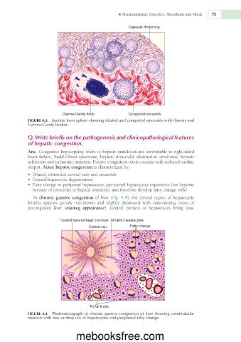
Capsular thickening
GamnaGandy body Congested sinusoids
FIGURE 4.3. Section from spleen showing dilated and congested sinusoids with fibrosis and
Gamna-Gandy bodies.
Q. Write briefly on the pathogenesis and clinicopathological features
of hepatic congestion.
Ans. Congestive hepatopathy refers to hepatic manifestations attributable to right-sided
heart failure, Budd-Chiari syndrome, hepatic sinusoidal obstruction syndrome, hepatic
infarction and ischaemic hepatitis. Passive congestion often coexists with reduced cardiac
output. Acute hepatic congestion is characterized by:
• Dilated, distended central vein and sinusoids
• Central hepatocyte degeneration
• Fatty change in periportal hepatocytes (periportal hepatocytes experience less hypoxia
because of proximity to hepatic arterioles, and therefore develop fatty change only)
In chronic passive congestion of liver (Fig. 4.4), the central region of hepatocytic
lobules appears grossly red–brown and slightly depressed with surrounding zones of
uncongested liver (nutmeg appearance). Central portion of hepatocytes being least
Central haemorrhagic necrosis Atrophic hepatocytes
Central vein Fatty change
Portal triads
FIGURE 4.4. Photomicrograph of chronic passive congestion of liver showing centrilobular
necrosis with loss or drop out of hepatocytes and peripheral fatty change.
mebooksfree.com

