
Page 1122 - Hematology_ Basic Principles and Practice ( PDFDrive )
P. 1122
988 Part VII Hematologic Malignancies
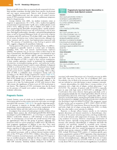
blueberry muffin lesions that are associated with congenital infection. TABLE Prognostically Important Genetic Abnormalities in
Skin nodules sometimes develop before bone marrow involvement 62.2 Pediatric Acute Myeloid Leukemia
and may regress spontaneously. Patients with elevated leukocyte
counts (hyperleukocytosis) may also present with central nervous Favorable
system (CNS) symptoms (seizure or stroke) or pulmonary symptoms t(8;21)(q22;q22)/RUNX1-RUNX1T1
related to hyperviscosity. inv(16)(p13.1;q22)/CBFβ-MYH11
Among children with AML, the median leukocyte count at t(16;16)(p13.1;q22)/CBFβ-MYH11
9
diagnosis is approximately 20 × 10 /L, with a median hemoglobin t(1;11)(q21;q23)/MLL-MLLT11
concentration of approximately 9 g/dL and a median platelet count NPM1/wt-FLT3
9
of 60 × 10 /L. Although about 10% of children with AML do not CEBPA
have morphologically detectable circulating blasts, careful examina- t(15;17)(q22;q12)/PML-RARα
tion of the peripheral blood smear will reveal leukemic cells in most Unfavorable
cases. Prolonged prothrombin, thrombin, and partial thromboplastin t(6;11)(q27;q23)/MLL-MLLT4
times, as well as decreased fibrinogen levels, are seen in the majority t(10;11)(p12;q23)/MLL-MLLT10
of patients with APL and about 5% of other AML cases. Abnormali- t(10;11)(p11.2;q23)/MLL-ABI1
ties of serum chemistries may include hyperuricemia, although this t(6;9)(p23;q34)/DEK-NUP214
is less common and less severe than hyperuricemia associated with t(8;16)(p11;p13)/MYST3-CREBBP
ALL. The presence of hypokalemia, a rare finding in patients with t(16;21)(q24;q22)/RUNX1-CBFA2T3
ALL, suggests a diagnosis of monoblastic leukemia. t(5;11)(q35;p15.5)/NUP98-NSD1
Among patients who present with circulating blasts, the differen- inv(16)(p13.3q24.3)/CBFA2T3-GLIS2
tial diagnosis is generally limited to various types of leukemia, FLT3-ITD
including AML, ALL, and juvenile myelomonocytic leukemia Monosomy 7
(JMML). For patients who do not have blasts in their blood at the Likely Unfavorable
time of presentation, the differential diagnosis is determined by signs IDH1, IDH2
and symptoms and may include aplastic anemia, autoimmune or RUNX1
inflammatory disease, infection, and solid malignancies. In most TET2
cases, the diagnosis of AML is made by bone marrow examination. DNMT3A
A bone marrow aspiration should be performed for morphologic Intermediate or Unknown
examination, immunophenotyping, genetic, and molecular studies,
whereas a bone marrow biopsy is used to assess cellularity. A diagnosis t(9;11)(p12;q23)/MLL-MLLT3
of AML is confirmed when 20% or more of nucleated bone marrow Other MLL
cells are blasts of myeloid origin or when the blasts contain AML- t(1;22)(p13;q13)/RBM15-MKL1
specific genetic lesions, regardless of blast percentage. When a diag-
nosis of AML is confirmed, most investigators classify each case
according to the World Health Organization criteria (Table 62.1).
Since AML may involve the CNS, examination of the cerebrospinal associated with normal karyotypes and a favorable outcome in adults
fluid (CSF) should also be performed as part of the workup of all with AML, but occur in less than 5% of childhood AML cases.
patients. Although CNS leukemia is traditionally defined as the Nevertheless, they are likely to be associated with a favorable outcome
presence of at least five leukocytes/µL of CSF with leukemic blast and are included as a low-risk feature in many treatment protocols.
cells present, the significance of lower levels of CNS involvement is Genetic abnormalities, for which there is strong evidence of an
not known. The diagnosis of CNS involvement may also be based association with a high risk of relapse, include monosomy 7 and
on the presence of cranial nerve palsies or radiologic evidence of FLT3-ITD. The outcome of patients with FLT3-ITD is especially
leukemic infiltration. poor in cases with high ratios of FLT3-ITD to wild-type FLT3.
Translocations that create chimeric fusion genes and likely confer a
poor prognosis include the t(6;11)(q27;q23)/MLL-MLLT4, t(10;11)
Prognostic Factors (p12;q23)/MLL-MLLT10, t(10;11)(p11.2;q23)/MLL-ABI1, t(5;11)
(q35;p15.5)/NUP98-NSD1, t(6;9)(p23;q34)/DEK-NUP214, t(8;16)
Genetic features, some of which can be identified by conventional (p11;p13)/MYST3-CREBBP, t(16;21)(q24;q22)/RUNX1-CBFA2T3,
karyotyping and others that require molecular techniques, are strongly and inv(16)(p13.3q24.3)/CBFA2T3-GLIS2. The prognostic impact
associated with outcome (Table 62.2). An equally important predic- of other lesions, such as mutations of WT1, IDH1, IDH2, RUNX1,
tor of outcome is response to therapy, which can be assessed by TET2, or DNMT3A, is not known. However, because they are associ-
morphologic, immunophenotypic, or molecular examination of the ated with a high risk of relapse among adults with AML, it is likely
bone marrow before and after each course of chemotherapy. that they also confer a poor outcome in children.
Investigators from almost all study groups consider children whose Response to therapy reflects features specific to the leukemia
leukemic blasts contain the t(8;21)(q22;q22)/RUNX1-RUNX1T1, (genetic alterations and inherent sensitivity to chemotherapy), char-
inv(16)(p13.1;q22)/CBFβ-MYH11, or t(16;16)(p13.1;q22)/CBFβ- acteristics of the patient (pharmacogenomics and drug metabolism),
MYH11 (collectively referred as CBF leukemia) to have low-risk as well as the intensity and components of therapy, and is therefore
AML. In contemporary clinical trials, the overall survival (OS) rates a key predictor of outcome. However, morphologic examination of
are approximately 90% for this group of patients. Although KIT the bone marrow, especially during periods of brisk hematopoietic
mutations confer an inferior prognosis in adults with CBF leukemia, recovery after intensive chemotherapy, is subjective and lacks the
their prognostic significance in children is not clear. Thus, most sensitivity and specificity required to accurately assess response.
clinical trials classify children with CBF leukemia as having low-risk Methods that rely on leukemia-specific features that distinguish
disease, regardless of other genetic abnormalities. residual leukemia cells from normal hematopoietic precursors can
Mutations of the NPM1 gene are seen primarily in AML cases provide more precise estimates of MRD. Techniques applicable to
with normal karyotypes, with or without internal tandem duplica- AML include RNA-based PCR analysis of leukemia-specific gene
tions of the FLT3 gene (FLT3-ITD). Children whose blasts contain fusions, quantitative analysis of WT1 expression, deep sequencing to
NPM1 mutations, normal karyotypes, and wild-type FLT3 appear to detect leukemia-specific mutations, and flow cytometric detection of
have an excellent prognosis, although the data to support their clas- aberrant immunophenotypes. Although PCR detection of fusion
sification as low-risk patients are not as strong as that for children transcripts is sensitive to a level of 0.01%–0.001%, it can be applied
with CBF leukemia. Similarly, biallelic mutations of CEBPA are to only about 50% of cases. In addition, the significance of persistence