
Page 1151 - Hematology_ Basic Principles and Practice ( PDFDrive )
P. 1151
1010 Part VII Hematologic Malignancies
HSCs from adult bone marrow, but hematopoiesis is sustained by
171
committed precursors. Most ETV6-RUNX1 leukemias show loss Nicastrin
of the normal ETV6 allele, suggesting that the leukemogenic effect
of ETV6-RUNX1 may be mediated in part by loss of wild-type ETV6
function. 172–175 Interestingly, germline loss-of-function ETV6 muta- Signaling Receiving
tions have been recently identified in patients with familial throm- cell γ-Secretase cell
bocytopenia and a predisposition to hematologic malignancies
including ALL, 176,177 further implicating wild-type ETV6 as an ALL Cleavage 2
tumor suppressor. Nucleus
RUNX1 is the DNA-binding component of the RUNX1-CBFβ NOTCH ICN
transcription factor complex disrupted by the t(8;21), t(3;21), and DSL ICN MAML
inv(16) in AML. RUNX1 is a transcription factor that is required for Cleavage 1 CSL
the expression of several hematopoietic genes involved in myeloid and
lymphoid development, including PU.1 and IL-3, although it can
178
also act as a transcriptional repressor in some settings. Homozygous Metalloprotease
disruption of the murine Runx1 or CBFB genes results in the lack of
definitive hematopoiesis, indicating that genes regulated by RUNX1
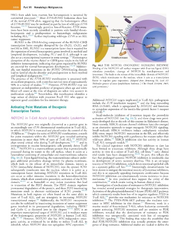
are essential for normal hematopoietic development. 179,180 Addition- Fig. 64.2 THE NOTCH1 ONCOGENIC SIGNALING PATHWAY.
ally, rare familial mutations in the RUNX1 DNA-binding domain Binding of the NOTCH1 cell surface receptor with δ serrate ligand (DSL)
lead to familial platelet disorder and predisposition to both myeloid stimulates proteolytic cleavage of NOTCH by metalloproteases and
and lymphoid malignancies. 177,181 γ-secretase. This leads to the release of the intracellular domain of NOTCH1
The presence of the ETV6-RUNX1 translocation is associated with (ICN), which translocates to the nucleus, where it acts as a transcription
an excellent prognosis, with event-free survival rates of approximately factor to regulate gene expression. (Adapted from Armstrong SA, Look AT:
90% in a variety of studies. 182–184 However, ETV6-RUNX1 may not Molecular genetics of acute lymphoblastic leukemia. J Clin Oncol 23:6306, 2005,
represent an independent predictor of prognosis when age and white with permission.)
blood cell count at the time of diagnosis are taken into account in
184
multivariate analysis. Nevertheless, this translocation identifies a
large subset of children with precursor B-cell ALL who appear to
represent good candidates for less intensive therapy. Additional NOTCH1 targets implicated in T-cell ALL pathogenesis
200
include the IL7R interleukin receptor, and the long noncoding
Activating Point Mutations of Oncogenic RNA LUNAR1, which is upregulated by NOTCH1 and functions
Transcription Factors to upregulate expression of the insulin-like growth factor 1 receptor
201
(IGF1R).
Small-molecule inhibitors of γ-secretase impair the proteolytic
NOTCH1 in T-Cell Acute Lymphoblastic Leukemia activation of NOTCH1 (see Fig. 64.2), and these drugs were previ-
ously developed due to the role of this enzyme in Alzheimer’s disease.
The NOTCH1 gene was originally discovered as a partner gene in More recently, SERCA calcium channel inhibitors have also emerged
an exceedingly rare t(7;9) chromosomal translocation in T-cell ALL, as an alternative approach to target oncogenic NOTCH1 signaling.
in which NOTCH1 is truncated and placed under the control of the Small-molecule SERCA inhibitors induce endoplasmic reticulum
185
TCRβ locus. Despite the rarity of NOTCH1 translocations, a search (ER) stress, impair NOTCH1 maturation in the ER, and effectively
for point mutations within the NOTCH1 gene revealed activating inhibit NOTCH1 signaling with a preference for mutant rather than
186
202
mutations in more than 50% of cases of T-cell ALL. NOTCH1 wild-type alleles. SERCA inhibition also has activity in human
plays several critical roles during T-cell development, 30,187–189 and its T-cell ALL xenograft models. 202
overexpression in murine hematopoietic cells potently drives T-cell The clinical experience with NOTCH1 inhibitors to date has
190
ALL. NOTCH1 is a transmembrane protein that is proteolytically been limited to γ-secretase inhibitors. Although these drugs have
186
processed during its transit to the cell surface, where it exists as a activity in vitro in a subset of T-cell ALL cell lines, early clinical
heterodimer consisting of extracellular and transmembrane subunits trial results have been disappointing. 203,204 In part, this reflects the
(Fig. 64.2). Upon ligand binding, the transmembrane subunit under- fact that prolonged systemic NOTCH inhibition is intolerable due
goes additional proteolytic cleavage within the plasma membrane, to development of severe secretory diarrhea. This is an on-target
which leads to the release of its intracellular domain, known as toxicity of NOTCH inhibition, which drives intestinal epithelial cells
ICN1 (intracellular domain of NOTCH1), into the cytosol. ICN1 to a secretory goblet cell fate. 205,206 Interestingly, dexamethasone has
subsequently translocates into the nucleus, where it is active as a been shown to mitigate the intestinal toxicity of γ-secretase inhibitors,
transcription factor. Activating NOTCH1 mutations in T-cell ALL and this is an especially appealing therapeutic combination because
can occur as either missense mutations in the heterodimerization NOTCH1 inhibition can simultaneously reverse resistance to dexa-
207
domain, which allow constitutive proteolytic activation of the ICN1 methasone. In vivo preclinical data support the utility of this
208
domain, 186,191 or as frameshift or stop codon mutations that lead combination, which awaits testing in human clinical trials.
to truncation of the PEST domain. The PEST domain regulates Investigation of mechanisms of resistance to NOTCH1 inhibition
proteasomal degradation of the protein, and these PEST-inactivating has revealed several potential strategies for therapeutic intervention.
mutations result in aberrant stabilization of ICN1 protein. 186,192 Activation of the phosphatidylinositol 3-kinase (PI3K)-AKT pathway,
Mutations in both regions are often found on the same allele in which commonly results from loss of its negative regulator PTEN in
209
cases of T-cell ALL, and are synergistic in increasing NOTCH1 T-cell ALL, provides one mechanism for resistance to NOTCH1
210
186
transcriptional output. Additionally, the NOTCH1 oncoprotein inhibition. The PTEN-PI3K-AKT pathway also mediates resis-
211
can also be stabilized by inactivating mutations of tumor suppressor tance to MYC inhibition in this disease. However, work in a
genes involved in its proteasomal degradation, including FBXW7 murine model of Kras-induced T-ALL, where leukemias commonly
and Cyclin C, as discussed later in this chapter. 193,194 MYC is an acquire activating NOTCH1 mutations and are sensitive to PI3K
important transcriptional target of NOTCH1, and it mediates many inhibitors, revealed that the development of resistance to PI3K
of the leukemogenic properties of NOTCH1 in human T-cell ALL inhibition was unexpectedly associated with loss of oncogenic
212
cells. 195–198 However, NOTCH1 also has MYC-independent onco- NOTCH1 signaling. This finding thus raises the possibility that
genic activity, as evidenced by its ability to induce T-cell ALL in dual PI3K-NOTCH1 inhibition may actually promote the emer-
199
zebrafish, where NOTCH1 does not upregulate MYC expression. gence of drug resistance. Additional studies are needed to define the