
Page 1174 - Hematology_ Basic Principles and Practice ( PDFDrive )
P. 1174
1022 Part VII Hematologic Malignancies
A C C E
B D D F
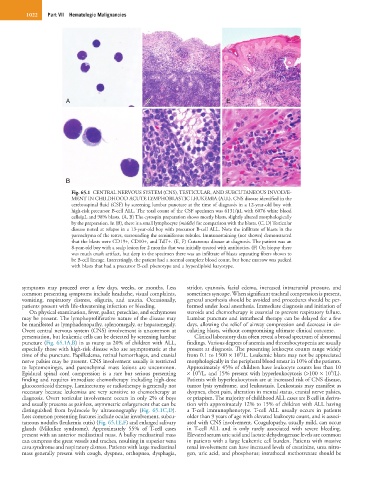
Fig. 65.1 CENTRAL NERVOUS SYSTEM (CNS), TESTICULAR, AND SUBCUTANEOUS INVOLVE-
MENT IN CHILDHOOD ACUTE LYMPHOBLASTIC LEUKEMIA (ALL). CNS disease identified in the
cerebrospinal fluid (CSF) by screening lumbar puncture at the time of diagnosis in a 12-year-old boy with
high-risk precursor B-cell ALL. The total count of the CSF specimen was 6131/µL with 6076 white blood
cells/µL and 98% blasts. (A, B) The cytospin preparation shows mostly blasts, slightly altered morphologically
by the preparation. In (B), there is a small lymphocyte (middle) for comparison with the blasts. (C, D) Testicular
disease noted at relapse in a 13-year-old boy with precursor B-cell ALL. Note the infiltrate of blasts in the
parenchyma of the testes, surrounding the seminiferous tubules. Immunostaining (not shown) demonstrated
that the blasts were CD19+, CD10+, and TdT+. (E, F) Cutaneous disease at diagnosis. The patient was an
8-year-old boy with a scalp lesion for 2 months that was initially treated with antibiotics. (F) On biopsy there
was much crush artifact, but deep in the specimen there was an infiltrate of blasts separating fibers shown to
be B-cell lineage. Interestingly, the patient had a normal complete blood count, but bone marrow was packed
with blasts that had a precursor B-cell phenotype and a hyperdiploid karyotype.
symptoms may proceed over a few days, weeks, or months. Less stridor, cyanosis, facial edema, increased intracranial pressure, and
common presenting symptoms include headache, visual complaints, sometimes syncope. When significant tracheal compression is present,
vomiting, respiratory distress, oliguria, and anuria. Occasionally, general anesthesia should be avoided and procedures should be per-
patients present with life-threatening infection or bleeding. formed under local anesthesia. Immediate diagnosis and initiation of
On physical examination, fever, pallor, petechiae, and ecchymoses steroids and chemotherapy is essential to prevent respiratory failure.
may be present. The lymphoproliferative nature of the disease may Lumbar puncture and intrathecal therapy can be delayed for a few
be manifested as lymphadenopathy, splenomegaly, or hepatomegaly. days, allowing the relief of airway compression and decrease in cir-
Overt central nervous system (CNS) involvement is uncommon at culating blasts, without compromising ultimate clinical outcome.
presentation, but leukemic cells can be detected by screening lumbar Clinical laboratory data often reveal a broad spectrum of abnormal
puncture (Fig. 65.1A,B) in as many as 20% of children with ALL, findings. Various degrees of anemia and thrombocytopenia are usually
especially those with high-risk disease who are asymptomatic at the present at diagnosis. The presenting leukocyte counts range widely
9
time of the puncture. Papilledema, retinal hemorrhages, and cranial from 0.1 to 1500 × 10 /L. Leukemic blasts may not be appreciated
nerve palsies may be present. CNS involvement usually is restricted morphologically in the peripheral blood smear in 10% of the patients.
to leptomeninges, and parenchymal mass lesions are uncommon. Approximately 45% of children have leukocyte counts less than 10
9
9
Epidural spinal cord compression is a rare but serious presenting × 10 /L, and 15% present with hyperleukocytosis (>100 × 10 /L).
finding and requires immediate chemotherapy including high-dose Patients with hyperleukocytosis are at increased risk of CNS disease,
glucocorticoid therapy. Laminectomy or radiotherapy is generally not tumor lysis syndrome, and leukostasis. Leukostasis may manifest as
necessary because leukemias are very sensitive to chemotherapy at dyspnea, chest pain, alteration in mental status, cranial nerve palsies,
diagnosis. Overt testicular involvement occurs in only 2% of boys or priapism. The majority of childhood ALL cases are B cell in deriva-
and usually presents as painless, asymmetric enlargement that can be tion with approximately 12% to 15% of children with ALL having
distinguished from hydrocele by ultrasonography (Fig. 65.1C,D). a T-cell immunophenotype. T-cell ALL usually occurs in patients
Less common presenting features include ocular involvement, subcu- older than 9 years of age with elevated leukocyte count, and is associ-
taneous nodules (leukemia cutis) (Fig. 65.1E,F) and enlarged salivary ated with CNS involvement. Coagulopathy, usually mild, can occur
glands (Mikulicz syndrome). Approximately 55% of T-cell cases in T-cell ALL and is only rarely associated with severe bleeding.
present with an anterior mediastinal mass. A bulky mediastinal mass Elevated serum uric acid and lactate dehydrogenase levels are common
can compress the great vessels and trachea, resulting in superior vena in patients with a large leukemic cell burden. Patients with massive
cava syndrome and respiratory distress. Patients with large mediastinal renal involvement can have increased levels of creatinine, urea nitro-
mass generally present with cough, dyspnea, orthopnea, dysphagia, gen, uric acid, and phosphorus; intrathecal methotrexate should be