
Page 1183 - Hematology_ Basic Principles and Practice ( PDFDrive )
P. 1183
Chapter 66 Acute Lymphoblastic Leukemia in Adults 1031
A C C D
B B E F G H
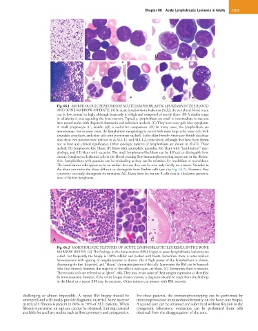
Fig. 66.1 MORPHOLOGIC FEATURES OF ACUTE LYMPHOBLASTIC LEUKEMIA IN THE BLOOD
AND BONE MARROW ASPIRATE. (A) In acute lymphoblastic leukemia (ALL), the peripheral blood count
can be low, normal or high, although frequently it is high and composed of mostly blasts. (B) A similar range
in cellularity is true regarding the bone marrow. Typically, lymphoblasts are small to intermediate in size and
have round nuclei with dispersed chromatin and indistinct nucleoli. (C) They have scant pale blue cytoplasm.
A small lymphocyte (C, middle, left) is useful for comparison. (D) In many cases, the lymphoblasts are
monotonous, but in some cases, the lymphoblast morphology is varied with some large cells, some cells with
abundant cytoplasm, and other cells with prominent nucleoli. In the older French–American–British classifica-
tion, these two patterns were referred to as ALL-L1 and ALL-L2, respectively, although they have been shown
not to have any clinical significance. Other cytologic variants of lymphoblasts are shown in (E–H). These
include (E) lymphocyte-like blasts, (F) blasts with azurophilic granules, (G) blasts with “hand-mirror” mor-
phology, and (H) blasts with vacuoles. The small lymphocyte-like blasts can be difficult to distinguish from
chronic lymphocytic leukemia cells in the blood, making flow immunophenotyping important in the distinc-
tion. Lymphoblasts with granules can be misleading as they can be mistaken for myeloblast or monoblasts.
The hand-mirror cells appear to be an artifact because they can be seen only focally on a smear. Vacuoles in
the blasts can make the blasts difficult to distinguish from Burkitt cells (see also Fig. 66.7). However, flow
cytometry can easily distinguish the immature ALL blasts from the mature B cells seen in a leukemic presenta-
tion of Burkitt lymphoma.
A B C
Fig. 66.2 MORPHOLOGIC FEATURES OF ACUTE LYMPHOBLASTIC LEUKEMIA IN THE BONE
MARROW BIOPSY. (A) The findings in the bone marrow (BM) biopsy in acute lymphoblastic leukemia are
varied, but frequently the biopsy is 100% cellular and packed with blasts. Sometimes there is some residual
hematopoiesis with sparing of megakaryocytes as shown. (B) A high power of the lymphoblasts is shown,
illustrating the fine, dispersed, and “blastic” chromatin pattern of the cells. Sometimes the BM can be hypocel-
lular (not shown); however, the majority of the cells in such cases are blasts. (C) Sometimes there is necrosis.
The necrotic cells are referred to as “ghost” cells. They may retain some of their antigen expression as identified
by immunostains; however, if the entire biopsy shows necrosis, a diagnosis should be made from the findings
in the blood or a repeat BM may be necessary. Other tumors can present with BM necrosis.
challenging or almost impossible. A repeat BM biopsy should be For these patients, the immunophenotyping can be performed by
attempted and will usually provide diagnostic material. Some increase immunoperoxidase immunohistochemistry on the bone core biopsy.
in reticulin fibrosis is present in 60% to 70% of ALL patients. When A second core can be obtained and submitted without fixation to the
fibrosis is extensive, an aspirate cannot be obtained, limiting material cytogenetic laboratory; evaluation can be performed from cells
available for ancillary studies such as flow cytometry and cytogenetics. obtained from the disaggregation of the core.