
Page 1184 - Hematology_ Basic Principles and Practice ( PDFDrive )
P. 1184
1032 Part VII Hematologic Malignancies
TABLE Antigens Used for Immunophenotyping of Acute
66.2 Lymphoblastic Leukemia a
Commonly Positive Variable Expression
B-ALL CD19 b CD20
cCD22 b CD34
cCD79a b CD45
Pax5 c CD13 d
CD10 CD33 d
sCD22 sIgM e
CD24 CD58 d
TdT CD38 d
T-ALL TdT CD1a
cCD3 f CD2
CD7 sCD3
A B CD4 g
CD5
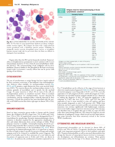
Fig. 66.3 CYTOCHEMISTRY IN ACUTE LYMPHOBLASTIC LEUKE- CD8 g
MIA. (A) The blasts in acute lymphoblastic leukemia are always myeloper- CD10
oxidase reaction negative. (B) Compare the blasts with a single positively CD34
reactive granulocyte with a black–blue reaction product. Evaluating for CD99
nonspecific esterase reactivity (α-naphthyl acetate esterase, or α-naphthyl CD19
butyrate esterase) might also be performed when the blasts are difficult to CD33 h
distinguish from monoblasts. CD79a
CD117 h
CD56
CD13 h
Organs other than the BM can be frequently involved. Extramed-
ullary or lymphomatous presentation is more common with T-acute a b Antigens are listed approximately in order of frequency.
lymphoblastic leukemia (T-ALL) than B-acute lymphoblastic leuke- c Almost always positive.
Most specific for B lineage but can be positive in t(8;21) acute myeloid
mia (B-ALL). The cytomorphology of the malignant cells in extra- leukemia.
medullary disease is similar to that described in the bone core biopsy. d Altered expression provides leukemia associated phenotype crucial for
Lymph node involvement is usually diffuse but can be partial with detection of minimal residual disease.
e
Rarely present.
sparing of the follicles. f Only marker considered lineage specific.
g Frequently coexpressed.
h Along with CD5 , CD1a-, CD8-, the expression of these antigens is helpful in
lo
CYTOCHEMISTRY identifying early T-cell precursor acute lymphoblastic leukemia. For prognostic
significance, please refer to discussion in text.
B-ALL, B-acute lymphoblastic leukemia; c, cytoplasmic; s, surface; T-ALL,
The use of cytochemistry to assign lineage has been largely replaced T-acute lymphoblastic leukemia.
by flow cytometry evaluation of the leukemic blast immunopheno-
type. However, when available, the myeloperoxidase reaction (Fig.
66.3A and B) permits a rapid distinction from acute myeloid leuke-
mia (AML). The reaction detects the myeloperoxidase enzyme in the B or T lymphoblasts can be reflective of the stage of development at
primary granules of myeloblasts and is specific for the myeloid which the transformation happened (Table 66.3). Of note, expression
lineage. An acute leukemia in which 3% or more of the cells are of myeloid antigens is seen frequently in B-ALL and T-ALL, as is the
myeloperoxidase positive is considered myeloid. There is excellent expression of T-cell antigens in B-ALL and B-cell antigens in T-ALL.
concordance between the myeloperoxidase reaction detected by Expression of individual myeloid antigens should not be a deterrent
cytochemistry and the myeloperoxidase molecule detected by flow to making the diagnosis of ALL. Of particular significance is the
cytometry. A block-like positivity has been described with the peri- subgroup of T-ALL that lack CD1a, CD8, and CD5 but show
odic acid-Schiff reaction that detects glycogen in almost 50% of ALL expression of one or more myeloid or stem cell markers and have
cases. been recently designated as early T-cell precursor ALL. While these
leukemias can have a gene expression profile and spectrum of genetic
mutations that overlaps significantly with biphenotypic leukemia,
IMMUNOPHENOTYPE most recent studies suggest improved outcomes using T-ALL therapy.
It is therefore best to recognize this type of ALL as a type of T-ALL
Based on large cooperative group studies in Europe and the United rather than mixed lineage leukemia. The criteria for diagnosis of acute
States, the incidence of B-ALL ranges from 75% to 80% and T-ALL leukemias of ambiguous lineage, which would include mixed pheno-
from 15% to 25%. B lymphoblasts cannot be distinguished from T type acute leukemia, have been extensively revised in the current
lymphoblasts by morphology. Extensive immunophenotypic charac- WHO classification.
terization is therefore required for the appropriate classification of
ALL and indeed distinction from certain subtypes of AML. When
adequate material is available, immunophenotyping should be per- CYTOGENETICS AND MOLECULAR GENETICS
formed using multicolor flow cytometry so that multiple antigens can
be detected simultaneously on the lymphoblasts (Fig. 66.4). When Chromosomal abnormalities can be detected in almost 80% of
interpreting the immunophenotypic data, it is important to remem- B-ALLs and 70% of T-ALLs. Cytogenetic classification remains the
ber that no single antigen is specific for any given lineage and multiple single most important prognostic factor in both pediatric and adult
antigens need to be evaluated to establish the correct diagnosis. The ALL. Numerical abnormalities as well as structural abnormalities that
panel of antibodies used for flow cytometry of a new leukemia and disrupt the function of transcription factors involved in hematopoi-
the pattern of expression seen in B-ALL and T-ALL are shown in etic development and differentiation are common. These genetic
Table 66.2. In addition, the combination of markers expressed on the abnormalities define the biology of the disease and have an impact