
Page 1248 - Hematology_ Basic Principles and Practice ( PDFDrive )
P. 1248
1094 Part VII Hematologic Malignancies
D
A B B CC E E
Fig. 68.6 PHOTOMICROGRAPH OF BONE MARROW BIOPSY OBTAINED FROM A PATIENT
WITH POLYCYTHEMIA VERA IN MYELOFIBROTIC PHASE DEMONSTRATING HYPERCELLU-
LARITY AND INCREASED NUMBER OF MEGAKARYOCYTES (×160).
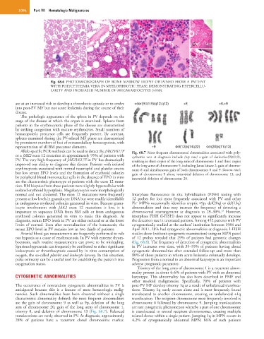
are at an increased risk to develop a thrombotic episode or to evolve +der(9)t(1;9)(q12;q12) +8 +8,+9
into post-PV MF but not acute leukemia during the course of their
disease.
The pathologic appearance of the spleen in PV depends on the
stage of the disease at which the organ is examined. Spleens from
patients in the erythrocytotic phase of the disease are characterized
by striking congestion with mature erythrocytes. Small numbers of
hematopoietic precursor cells are frequently present. By contrast,
spleens examined during the PV-related MF phase are characterized
by prominent numbers of foci of extramedullary hematopoiesis, with
representation of all BM precursor elements. +9 del(13)(q14q22) del(20)(q11q13)
Allele-specific PCR methods can be used to detect the JAK2V617F Fig. 68.7 Most frequent chromosomal abnormalities associated with poly-
or a JAK2 exon 12 mutation in approximately 95% of patients with cythemia vera at diagnosis include (top row) a gain of derivative(9)t(1;9),
PV. The very high frequency of JAK2V617F in PV has dramatically resulting in three copies of the long arms of chromosome 1 and three copies
improved our ability to diagnose this disease. Patients with isolated of the long arms of chromosome 9, including Janus kinase 2; gain of chromo-
erythrocytosis associated with normal neutrophil and platelet counts some 8 and simultaneous gain of both chromosomes 8 and 9 (bottom row);
but low serum EPO levels and the formation of erythroid colonies gain of chromosome 9 alone; interstitial deletion of chromosome 13; and
by peripheral blood mononuclear cells in the absence of EPO in vitro interstitial deletion of chromosome 20.
are the characteristic phenotype of patients with the exon 12 muta-
tion. BM biopsies from these patients were slightly hypercellular with
isolated erythroid hyperplasia. Megakaryocytes were morphologically
normal and not clustered. The exon 12 mutations were frequently Interphase fluorescence in situ hybridization (FISH) testing with
present at low levels in granulocyte DNA but were readily identifiable 12 probes for loci most frequently associated with PV and other
−
in endogenous erythroid colonies generated in vitro. Because granu- Ph MPNs occasionally identifies cryptic +9p, del(20q) or del(13q)
locyte involvement with JAK2 exon 12 mutations is low, it is abnormalities and thus may increase the frequency of detecting a
23
important to sequence DNA from BM cells or from endogenous chromosomal rearrangement at diagnosis to 29–30%. However,
erythroid colonies generated in vitro to make this diagnosis. At interphase FISH (I-FISH) does not appear to significantly increase
diagnosis, serum EPO levels in PV are either reduced or at the lower the detection rate in untreated patients. Among 452 patients with PV
limits of normal. Even after normalization of the hematocrit, the cytogenetically studied at the authors’ institution between 1984 and
serum EPO level in PV remains low in two thirds of patients. April 2011, 28% had cytogenetic abnormalities at diagnosis. I-FISH
Arterial blood gas measurements are frequently performed to rule studies alone (without cytogenetic examination) using an MPN panel
out hypoxia as a cause of erythrocytosis. In PV with extreme throm- of 12 probes revealed that 29% of patients had genomic changes
bocytosis, such routine measurements can prove to be misleading. (Fig. 68.8). The frequency of detection of cytogenetic abnormalities
Spurious hypoxemia can frequently be attributed to either significant in PV increases over time, with 35–55% of patients having clonal
leukocytosis or thrombocytosis caused by in vitro consumption of cytogenetic abnormalities after extended follow-up and more than
oxygen, the so-called platelet and leukocyte larceny. In this situation, 80% of those patients in whom acute leukemia eventually develops.
pulse oximetry can be a useful tool for establishing the patient’s true Progression from a normal to an abnormal karyotype is an important
oxygenation status. adverse prognostic parameter.
Trisomy of the long arms of chromosome 1 is a recurrent abnor-
mality present in about 4–6% of patients with PV with an abnormal
CYTOGENETIC ABNORMALITIES karyotype. This abnormality has also been described in PMF and
other myeloid malignancies. Specifically, 70% of patients with
The occurrence of nonrandom cytogenetic abnormalities in PV is post-PV MF develop trisomy 1q as a result of unbalanced transloca-
anticipated because this is a feature of most hematologic malig- tions. Trisomy 1q rarely occurs alone and is most frequently found
nancies. Such abnormalities have been observed without a single translocated to another chromosome, creating an unbalanced +1q
characteristic abnormality defined; the most frequent abnormalities translocation. The recipient chromosome most frequently involved is
are the gain of chromosome 9 as well as 9p, deletion of the long chromosome 6 followed by chromosome 9. Jumping translocations
arm of chromosome 20, gain of the long arms of chromosome 1, are rare cytogenetic phenomenon whereby a part of one chromosome
trisomy 8, and deletion of chromosome 13 (Fig. 68.7). Balanced is translocated to several recipient chromosomes, creating multiple
translocations are rarely observed in PV. At diagnosis, approximately related clones within a single patient. Jumping 1q in MPN occurs in
28% of patients have a recurrent clonal chromosome marker. 4.2% of cytogenetically abnormal MPN; 86% of such patients