
Page 1250 - Hematology_ Basic Principles and Practice ( PDFDrive )
P. 1250
1096 Part VII Hematologic Malignancies
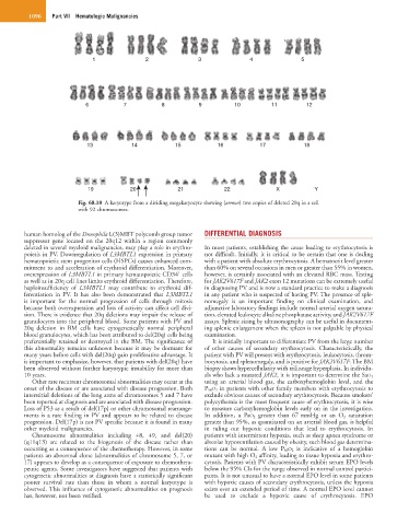
1 2 3 4 5
6 7 8 9 10 11 12
13 14 15 16 17 18
19 20 21 22 X Y
Fig. 68.10 A karyotype from a dividing megakaryocyte showing (arrows) two copies of deleted 20q in a cell
with 92 chromosomes.
human homolog of the Drosophila L(3)MBT polycomb group tumor DIFFERENTIAL DIAGNOSIS
suppressor gene located on the 20q12 within a region commonly
deleted in several myeloid malignancies, may play a role in erythro- In most patients, establishing the cause leading to erythrocytosis is
poiesis in PV. Downregulation of L3MBTL1 expression in primary not difficult. Initially, it is critical to be certain that one is dealing
hematopoietic stem progenitor cells (HSPCs) causes enhanced com- with a patient with absolute erythrocytosis. A hematocrit level greater
mitment to and acceleration of erythroid differentiation. Moreover, than 60% on several occasions in men or greater than 55% in women,
+
overexpression of L3MBTL1 in primary hematopoietic CD34 cells however, is certainly associated with an elevated RBC mass. Testing
as well as in 20q cell lines limits erythroid differentiation. Therefore, for JAK2V617F and JAK2 exon 12 mutations can be extremely useful
haploinsufficiency of L3MBTL1 may contribute to erythroid dif- in diagnosing PV and is now a standard practice to make a diagnosis
ferentiation in PV. It has also been demonstrated that L3MBTL1 in any patient who is suspected of having PV. The presence of sple-
is important for the normal progression of cells through mitosis nomegaly is an important finding on clinical examination, and
because both overexpression and loss of activity can affect cell divi- adjunctive laboratory findings include normal arterial oxygen satura-
sion. There is evidence that 20q deletions may impair the release of tion, elevated leukocyte alkaline phosphatase activity, and JAK2V617F
granulocytes into the peripheral blood. Some patients with PV and assays. Splenic sizing by ultrasonography can be useful in document-
20q deletion in BM cells have cytogenetically normal peripheral ing splenic enlargement when the spleen is not palpable by physical
blood granulocytes, which has been attributed to del(20q) cells being examination.
preferentially retained or destroyed in the BM. The significance of It is initially important to differentiate PV from the large number
this abnormality remains unknown because it may be dormant for of other causes of secondary erythrocytosis. Characteristically, the
many years before cells with del(20q) gain proliferative advantage. It patient with PV will present with erythrocytosis, leukocytosis, throm-
is important to emphasize, however, that patients with del(20q) have bocytosis, and splenomegaly, and is positive for JAK2V617F. The BM
been observed without further karyotypic instability for more than biopsy shows hypercellularity with trilineage hyperplasia. In individu-
10 years. als who lack a mutated JAK2, it is important to determine the SaO 2
Other rare recurrent chromosomal abnormalities may occur at the using an arterial blood gas, the carboxyhemoglobin level, and the
onset of the disease or are associated with disease progression. Both P 50 O 2 in patients with other family members with erythrocytosis to
interstitial deletions of the long arms of chromosomes 5 and 7 have exclude obvious causes of secondary erythrocytosis. Because smokers’
been reported at diagnosis and are associated with disease progression. polycythemia is the most frequent cause of erythrocytosis, it is wise
Loss of P53 as a result of del(17p) or other chromosomal rearrange- to measure carboxyhemoglobin levels early on in the investigation.
ments is a rare finding in PV and appears to be related to disease In addition, a PaO 2 greater than 67 mmHg or an O 2 saturation
progression. Del(17p) is not PV specific because it is found in many greater than 95%, as quantitated on an arterial blood gas, is helpful
other myeloid malignancies. in ruling out hypoxic conditions that lead to erythrocytosis. In
Chromosome abnormalities including +8, +9, and del(20) patients with intermittent hypoxia, such as sleep apnea syndrome or
(q11q13) are related to the biogenesis of the disease rather than alveolar hypoventilation caused by obesity, such blood gas determina-
occurring as a consequence of the chemotherapy. However, in some tions can be normal. A low P 50 O 2 is indicative of a hemoglobin
patients an abnormal clone (abnormalities of chromosome 5, 7, or mutant with high O 2 affinity, leading to tissue hypoxia and erythro-
17) appears to develop as a consequence of exposure to chemothera- cytosis. Patients with PV characteristically exhibit serum EPO levels
peutic agents. Some investigators have suggested that patients with below the 95% CIs for the range observed in normal control partici-
cytogenetic abnormalities at diagnosis have a statistically significant pants. It is not unusual to have a normal EPO level in some patients
poorer survival rate than those in whom a normal karyotype is with hypoxic causes of secondary erythrocytosis, unless the hypoxia
observed. This influence of cytogenetic abnormalities on prognosis exists over an extended period of time. A normal EPO level cannot
has, however, not been verified. be used to exclude a hypoxic cause of erythrocytosis. EPO