
Page 1280 - Hematology_ Basic Principles and Practice ( PDFDrive )
P. 1280
1126 Part VII Hematologic Malignancies
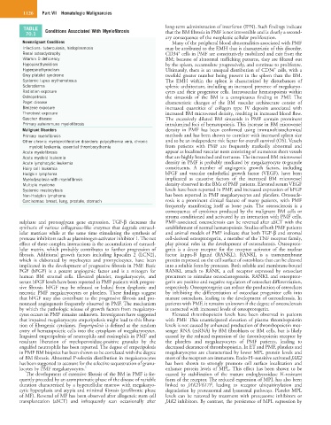
TABLE Conditions Associated With Myelofibrosis long-term administration of interferon (IFN). Such findings indicate
70.1 that the BM fibrosis in PMF is not irreversible and is clearly a second-
ary consequence of the neoplastic cellular proliferation.
Nonmalignant Conditions Many of the peripheral blood abnormalities associated with PMF
Infections: tuberculosis, histoplasmosis may be attributed to the EMH that is characteristic of this disorder.
+
Renal osteodystrophy CD34 cells in PMF are constitutively mobilized and exit from the
Vitamin D deficiency BM; because of abnormal trafficking patterns, they are filtered out
Hypoparathyroidism by the spleen, accumulate progressively, and continue to proliferate.
+
Hyperparathyroidism Ultimately, there is an unequal distribution of CD34 cells, with a
Gray platelet syndrome twofold greater number being present in the spleen than the BM.
Systemic lupus erythematosus The EMH within the spleen is characterized by disturbances of
Scleroderma splenic architecture, including an increased presence of megakaryo-
Radiation exposure cytes and their progenitor cells. Intravascular hematopoiesis within
Osteopetrosis the sinusoids of the BM is a conspicuous finding in PMF. The
Paget disease characteristic changes of the BM vascular architecture consist of
Benzene exposure increased quantities of collagen type IV deposits associated with
Thorotrast exposure increased BM microvessel density, resulting in increased blood flow.
Gaucher disease The excessively dilated BM sinusoids in PMF contain prominent
Primary autoimmune myelofibrosis intraluminal foci of hematopoiesis. This increase in BM microvessel
Malignant Disorders density in PMF has been confirmed using immunohistochemical
Primary myelofibrosis methods and has been shown to correlate with increased spleen size
Other chronic myeloproliferative disorders: polycythemia vera, chronic and to be an independent risk factor for overall survival (OS). Vessels
myeloid leukemia, essential thrombocythemia from patients with PMF are frequently markedly abnormal and
Acute myelofibrosis appear as localized vascular nests consisting of numerous short vessels
Acute myeloid leukemia that are highly branched and tortuous. The increased BM microvessel
Acute lymphocytic leukemia density in PMF is probably mediated by megakaryocyte α-granule
Hairy cell leukemia constituents. A number of angiogenic growth factors, including
Hodgkin lymphoma bFGF and vascular endothelial growth factor (VEGF), have been
Myelodysplasia with myelofibrosis implicated as causative factors of the increased BM microvessel
Multiple myeloma density observed in the BMs of PMF patients. Elevated serum VEGF
Systemic mastocytosis levels have been reported in PMF, and increased expression of bFGF
Non-Hodgkin lymphoma has been reported in PMF megakaryocytes and platelets. Osteoscle-
Carcinomas: breast, lung, prostate, stomach rosis is a prominent clinical feature of many patients, with PMF
frequently manifesting itself as bone pain. The osteosclerosis is a
consequence of cytokines produced by the malignant BM cells or
stroma conditioned and activated by an interaction with PMF cells.
sulphate and proteoglycan gene expression. TGF-β decreases the PMF-associated osteosclerosis can be reversed after aSCT with the
synthesis of various collagenase-like enzymes that degrade extracel- establishment of normal hematopoiesis. Studies of both PMF patients
lular matrices while at the same time stimulating the synthesis of and animal models of PMF indicate that both TGF-β and stromal
protease inhibitors such as plasminogen activator inhibitor 1. The net cell-derived osteoprotegerin, a member of the TNF receptor family,
effect of these complex interactions is the accumulation of extracel- play pivotal roles in the development of osteosclerosis. Osteoprote-
lular matrix, which probably contributes to further progression of gerin is a decoy receptor for the receptor activator of the nuclear
fibrosis. Additional growth factors including lipocalin 2 (LCN2), factor kappa-B ligand (RANKL). RANKL is a transmembrane
which is elaborated by myelocytes and promyelocytes, have been protein expressed on the cell surface of osteoblasts that can be cleaved
implicated in the development of progressive fibrosis in PMF. Basic into a soluble form by proteases. Both soluble and membrane-bound
FGF (bFGF) is a potent angiogenic factor and is a mitogen for RANKL attach to RANK, a cell receptor expressed by osteoclast
human BM stromal cells. Elevated platelet, megakaryocyte, and precursors to stimulate osteoclastogenesis. RANKL and osteoprote-
serum bFGF levels have been reported in PMF patients with progres- gerin are positive and negative regulators of osteoclast differentiation,
sive fibrosis. bFGF may be released or leaked from dysplastic and respectively. Osteoprotegerin can reduce the production of osteoclasts
necrotic PMF megakaryocytes or platelets. These findings suggest by inhibiting the differentiation of osteoclast progenitor cells into
that bFGF may also contribute to the progressive fibrosis and pro- mature osteoclasts, leading to the development of osteosclerosis. In
nounced angiogenesis frequently observed in PMF. The mechanism patients with PMF, it remains unknown if the degree of osteosclerosis
by which the pathologic release of growth factors from megakaryo- is corrected with increased levels of osteoprotegerin.
cytes occurs in PMF remains unknown. Investigators have suggested Elevated thrombopoietin levels have been observed in patients
that impaired megakaryocyte emperipolesis might lead to this libera- with PMF. This unanticipated elevation of plasma thrombopoietin
tion of fibrogenic cytokines. Emperipolesis is defined as the random levels is not caused by enhanced production of thrombopoietin mes-
entry of hematopoietic cells into the cytoplasm of megakaryocytes. senger RNA (mRNA) by BM fibroblasts or BM cells, but is likely
Impaired emperipolesis of neutrophils and eosinophils in PMF and caused by the reduced expression of the thrombopoietin receptor by
resultant liberation of myeloperoxidase-positive granules by the the platelets and megakaryocytes of PMF patients, leading to
engulfed neutrophils has been reported. The degree of emperipolesis decreased clearance of thrombopoietin. In ET and PMF, platelets and
in PMF BM biopsies has been shown to be correlated with the degree megakaryocytes are characterized by lower MPL protein levels and
of BM fibrosis. Abnormal P-selectin distribution in megakaryocytes most of the receptors are immature. Endo-H–sensitive activated JAK2
has been suggested to account for the selective sequestration of granu- has been shown to strongly promote cell surface localization and
locytes by PMF megakaryocytes. 3 enhance protein levels of MPL. This effect has been shown to be
The development of extensive fibrosis of the BM in PMF is fre- caused by stabilization of the mature endoglycosidase H-resistant
quently preceded by an asymptomatic phase of the disease of variable form of the receptor. The reduced expression of MPL has also been
duration characterized by a hypercellular marrow with megakaryo- linked to JAK2V617F, leading to receptor ubiquitinylation and
cytic hyperplasia and atypia and minimal fibrosis (prefibrotic phase degradation by proteasomal and lysosomal pathways. Platelet MPL
of MF). Reversal of MF has been observed after allogeneic stem cell levels can be restored by treatment with proteasome inhibitors or
transplantation (aSCT) and infrequently seen occasionally after JAK2 inhibitors. By contrast, the persistence of MPL expression by