
Page 1307 - Hematology_ Basic Principles and Practice ( PDFDrive )
P. 1307
Chapter 71 Eosinophilia, Eosinophil-Associated Diseases, Eosinophilic Leukemias, and the Hypereosinophilic Syndromes 1153
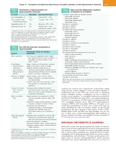
TABLE Classification of Hypereosinophilia and TABLE Organ-restricted (Inflammatory) Conditions
71.3 Hypereosinophilic Syndromes 71.4B Accompanied by Eosinophilia
HE Variant Abbreviation Associated HES Variant Eosinophilic gastrointestinal disorders (EGIDs)
Eosinophilic esophagitis
Familial/hereditary HE HE FA Familial HES = HES F
Eosinophilic gastritis
HE of undetermined/ HE US a Idiopathic HES = HES I a Eosinophilic gastroenteritis
Unknown Significance Eosinophilic colitis
Neoplastic/primary HE HE N Neoplastic HES = HES N Eosinophilic pancreatitis
Reactive/secondary HE HE R Reactive HES = HES R Eosinophilic hepatitis
b Eosinophilic ascites
Lymphocytic variant = HES L
Pulmonary eosinophilic syndromes
a In patients with HE US , the presence of HES is excluded by definition; as soon
as HES is diagnosed in a patient with HE US , the diagnosis changes to HES I , Eosinophilic asthma
unless an underlying etiology (condition/disease) is also found. Eosinophilic bronchitis
b HES L is regarded a special variant of HES R . Eosinophilic pneumonia
HE, Hypereosinophilia; HES, hypereosinophilic syndrome; HES F , familial HES; Eosinophil pleuritis
HES I , idiopathic HES; HES L , lymphocytic variant HES; HES N , primary Eosinophilic nephritis
(neoplastic) HES; HES R , reactive HES; HE US , hypereosinophilia of undetermined
(unknown) clinical significance. Eosinophilic cystitis
Eosinophilic endometritis and myometritis
Eosinophilic mastitis
TABLE Rare HES-Like Syndromes Accompanied by Eosinophilic ocular disorders
71.4A Hypereosinophilia Eosinophilic myocarditis
Eosinophilic synovitis
Characteristic Features and Laboratory Skin diseases/conditions associated with eosinophilia a
Syndrome Abnormalities
Allergic contact dermatitis
Gleich syndrome Cyclic recurrent angioedema, fever, weight Atopic dermatitis
gain, elevated polyclonal IgM, increased IL-5 Drug reactions
production, evidence of clonal T cells Bullous pemphigoid and pemphigoid variants
(CD3 T cells) a Eosinophilic cellulitis (Wells syndrome)
–
Churg-Strauss Stage I: Asthma and rhinosinusitis (isolated) Radiotherapy-related eruptions associated with eosinophilia
syndrome (CSS) = Stage II: Eosinophilic phase (HES-like) Eosinophilic pustular folliculitis
eosinophilic Stage III: Vasculitic phase defined by Infestations (parasitic) and infections (bacterial, viral, fungal)
granulomatosis necrotizing (small-vessel) vasculitis and HE involving the skin
with polyangiitis with pulmonary infiltrates and/or neuropathy Mycosis fungoides
(EGPA) (ANCA+ and ANCA− subvariants). Pachydermatous eosinophilic dermatitis
Kimura disease (skin and lymph nodes)
Eosinophilia myalgia Severe myalgia ± fever (flu-like), edema, Eosinophilic panniculitis
syndrome (EMS) fatigue, weight loss, neurologic symptoms,
skin abnormalities a Many other skin abnormalities can be accompanied by eosinophilia, especially
Epidemic cases have been reported to result when a systemic inflammatory disease process is present and triggers
eosinophilia.
from exposure to L-tryptophan (toxic oil
syndrome).
Eosinophilic fasciitis Scleroderma-like condition with painful emphasize the need for more comprehensive epidemiologic studies
(EF) = Shulman swelling of the skin and chronic induration using currently available diagnostic criteria and robust registries. A
syndrome (limbs and trunk) as well as laboratory signs complicating issue is that HES may coexist with an underlying
of systemic inflammation primary eosinophil disorder, a lymphoma, but also an allergy or
3
Omenn syndrome Severe combined immunodeficiency (SCID) another reactive condition. In eosinophilic leukemia presenting with
(OS) with well-populated lymphatic organs (OS the FIP1L1-PDGFRA fusion gene (F/P), a clear male predominance
paradox) is found. As a consequence, any HES registry including patients with
Autosomal recessive genetic (pediatric) disease F/P+ eosinophilic leukemia and/or other F/P+ myeloproliferative
(recurrent mutations in RAG1 or RAG2) neoplasms (MPNs) with eosinophilia (MPN-eo) will report on an
Clinical findings: erythroderma, overall male predominance of HES, even if other HES types are
hepatosplenomegaly, lymphadenopathy, evenly distributed in males and females. Indeed, no clear gender
increased IgE, diarrhea, weight loss predominance has been reported in other groups of HE or HES.
(GVHD-like condition with autoreactive T Familial forms of HE and HES have been described, but are very
cells) rare. In these cases, a hyper-immunoglobulin (Ig)E syndrome or
Hyper-IgE syndrome Hereditary immunodeficiency syndrome with Omenn syndrome (OS) have to be excluded. No cases of familial F/
(HIES) elevated IgE, recurrent severe infections, P+ leukemia have been reported to date.
often with skin eczema and facial anomalies
Known recurrent gene mutations: MORPHOLOGY AND PHENOTYPE OF EOSINOPHILS
Autosomal dominant variant: STAT3 mutations
Autosomal recessive variant: DOCK8 mutations
PGM3 mutations Eosinophils contain three granule populations, namely (1) electron-
dense, round, primary progranules present mainly at the promyelocyte
a EGPA/CSS with clonal T cells is often regarded as special variant of lymphoid and myelocyte stages, (2) specific (secondary) eosinophilic granules
variant HES (HES L).
ANCA, Anti-neutrophil cytoplasmic antibodies; DOCK8, dedicator of cytokinesis with an electron-dense crystalloid core, specifically found in mature
8; GVHD, graft-versus-host disease; HE, hypereosinophilia; HES, eosinophils, and (3) small-sized granules, which may contain catalase,
1
hypereosinophilic syndrome; IgE, immunoglobulin E; PGM3, acid phosphatase, and arylsulfatase. The large, specific granule is the
phosphoglucomutase 3; RAG, recombination-activating gene; STAT3, signal major source of cytotoxic and proinflammatory cationic proteins.
transducer and activator of transcription-3.
Eosinophils also contain lipid bodies that may serve as a source of