
Page 1368 - Hematology_ Basic Principles and Practice ( PDFDrive )
P. 1368
1214 Part VII Hematologic Malignancies
A B
C D
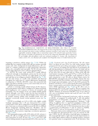
Fig. 75.2 MORPHOLOGIC VARIATIONS OF REED-STERNBERG (RS) CELLS IN CLASSIC
HODGKIN LYMPHOMA (cHL). The diagnostic RS cells (A) are large binucleated (arrow) or multinucleated
cells (arrowhead) with distinct nuclear membrane, prominent nucleolus in each nuclear lobe, and abundant
amphophilic cytoplasm. Hodgkin cells are mononuclear variants of RS cells with similar cytonuclear features
(B); numerous eosinophils and granulocytes are noted in the background. Mummified cells are degenerated
RS and Hodgkin cells with pyknotic nuclei and condensed cytoplasm (C). Lacunar cells, characteristic of
nodular sclerosis cHL, have abundant pale cytoplasm which frequently retracts in formalin fixed tissue (D).
imparting a prominent nodular pattern (Fig. 75.3A). Within the 75.4B). Consistent with their B-cell derivation, RS cells express
nodules there are a variable number of RS cells and variants, especially PAX-5 in almost all cases (95%) but with weaker intensity when
lacunar cells, with a background of mixed inflammatory cells com- compared with the surrounding nonneoplastic small B cells (see Fig.
posed of a variable proportion of small lymphocytes, histiocytes, 75.4C). However, in keeping with their defective B-cell program, RS
plasma cells, eosinophils, and neutrophils. The RS cells and variants cells lack Ig production as evidenced by absence of J chain, and are
may be singly dispersed or form confluent aggregates/sheets. negative for most other B cell−associated antigens: CD20 (expressed
In MCCHL the lymph node architecture is usually diffusely in only 20%−30% of cases; often only in a subset of RS cells with
obliterated, although an interfollicular pattern may be seen in early weak/variable intensity), CD19, and CD79a; as well as B-cell tran-
involvement. In contrast to NSCHL, the nodal capsule is not thick- scriptional factors OCT-2 and BOB.1 (each expressed in 10% of
ened and there are no collagenous bands of fibrosis (see Fig. 75.3B). cases; coexpression is rare). RS cells are almost always positive for
RS cells and variants are usually easily identified and dispersed IRF4/MUM1 and negative for CD45 and EMA, features that may
throughout the nodal tissue in a mixed inflammatory background. help distinguish cHL from NLPHL. Expression of other hematopoi-
In comparison to NSCHL, MCCHL is more often associated with etic lineage−associated markers, such as T cells (CD4, granzyme B),
higher stage disease and EBV positivity, and is more likely seen in the dendritic cells (fascin, CCL17), and myeloid cells (colony stimulating
HIV-infected patient population. factor 1 receptor and α 1 -antitrypsin) is also often present. EBV LMP1
LRCHL is a relatively recently defined subtype of cHL character- and/or EBER expression (see Fig. 75.4D) by RS cells is seen in
ized by the presence of RS cells in a background of almost exclusively approximately 40% of cHL cases overall in Western countries, but
small lymphocytes, with a paucity or absence of eosinophils and mostly in MCCHL and LDCHL and less frequently in NSCHL and
neutrophils. The vast majority of the cases exhibit nodular growth LRCHL. However, an association with EBV is seen in up to 90% of
pattern, although a rare diffuse variant has also been described. In cHL cases in developing countries and nearly all cases in the HIV
the vast majority of the cases the affected lymph node is obliterated patient population.
by multiple expansile nodules with expanded mantle zones and The nonneoplastic background lymphocytes, with the exception
regressed, eccentrically located residual germinal centers (see Fig. of LRCHL, are composed of predominantly T cells with marked
75.3C). predominance of CD4-positive cells that coexpress CD25 and
LDCHL is exceedingly rare (<1% of cHL) with a highly variable FOXP3, consistent with immunosuppressive regulatory T-cells
histologic appearance, but in all cases characterized by a relative (TReg). In addition there is a significant population of TH2 cells.
predominance of RS cells in comparison to the background lympho- TReg and TH2 cells are attracted by cytokines (CCL5, CCL17, and
cytes. Some cases are characterized by scattered RS cells in a diffusely CCL22) secreted by RS cells. In HIV-infected patients there is often
fibrotic background containing histiocytes, fibroblasts, and few a predominance of CD8-positive T cells. In contrast to NLPHL,
lymphocytes. In others, sheets of bizarre, pleomorphic, or anaplastic- CD57-positive T cells are not increased in number in cHL.
appearing RS cells are present, imparting a sarcomatous appearance Polymerase chain reaction studies performed on RS cells procured
(see Fig. 75.3D). by microdissection demonstrated that in the vast majority of cHL
The immunophenotypic profile of RS cells in all cHL subtypes is cases (>98%), the RS cells harbor clonal IgH gene rearrangement.
similar. RS cells are strongly positive for CD30 with membranous The rearranged IgH shows a high load of somatic hypermutation in
and Golgi pattern in nearly all cases (Fig. 75.4A), and CD15 with the variable region without evidence of ongoing mutation, consistent
variable staining intensity in approximately 80% of the cases (see Fig. with germinal center or postgerminal center B-cell derivation. Rare