
Page 1369 - Hematology_ Basic Principles and Practice ( PDFDrive )
P. 1369
Chapter 75 Hodgkin Lymphoma 1215
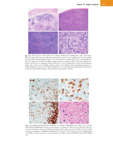
A B
C D
Fig. 75.3 HISTOLOGIC PATTERNS OF CLASSIC HODGKIN LYMPHOMA (cHL) SUBTYPES.
Nodular sclerosis cHL characteristically shows multinodular pattern on low magnification, where the nodules
are surrounded by broad collagenous bands. The nodal capsule is markedly sclerotic (A). In mixed cellularity
cHL, the lymph node architecture is diffusely obliterated by the neoplastic infiltrate. The nodal capsule is not
sclerotic and there are no collagenous bands of fibrosis (B). A case of lymphocyte-rich cHL where the affected
lymph node is obliterated by multiple expansile nodules, with strikingly similar morphologic appearance to
nodular lymphocyte predominant Hodgkin lymphoma (C). A case of lymphocyte-depleted cHL with presence
of numerous pleomorphic/anaplastic appearing Reed-Sternberg (RS) cells (D).
A B
C D
Fig. 75.4 IMMUNOPHENOTYPIC PROFILE OF REED-STERNBERG (RS) CELLS IN CLASSIC
HODGKIN LYMPHOMA. The RS cells and variants are positive for CD15 (A) and CD30 (B), with char-
acteristic membranous staining and Golgi accentuation. These cells are positive for PAX-5, but with weaker
intensity in comparison to background small reactive B cells (C). The RS cells are positive for Epstein-Barr
virus (EBV) as demonstrated by in situ hybridization for EBV-encoded RNA/EBV-encoded RNA (EBER)
(D).