
Page 1370 - Hematology_ Basic Principles and Practice ( PDFDrive )
P. 1370
1216 Part VII Hematologic Malignancies
A B
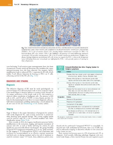
Fig. 75.5 IMMUNOHISTOCHEMICAL ANALYSIS OF PDL-1 EXPRESSION IN CLASSIC HODGKIN
LYMPHOMA (cHL) AND NODULAR LYMPHOCYTE-PREDOMINANT HODGKIN LYMPHOMA
(NLPHL). (A) A case of nodular sclerosis cHL showing distinct membranous expression for PDL-1 by
Reed-Sternberg (RS) cells (arrows). PDL-1 also highlights cell processes of tumor-infiltrating histiocytes
(arrowheads) with granular staining pattern. (B) A case of nodular lymphocyte predominant Hodgkin lym-
phoma showing lymphocyte predominant (LP) cells (arrows) are negative for PDL-1, whereas the background
tumor-infiltrating histiocytes (arrowheads) are highlighted by PDL-1 with granular pattern of staining by
their cell processes.
cases harboring T-cell receptor gene rearrangements have also been TABLE Cotswold-Modified Ann Arbor Staging System for
documented. Genetic structural alterations that modulate the tumor 75.2 Hodgkin Lymphoma
microenvironment have also been observed. For example, 9p24.1
amplification leads to enhanced PDL-1 expression by RS cells, which Stage Criteria
inhibit T-cell effector functions by binding to PD-1 on T cells, I Disease affecting a single lymph node region or lymphoid
enhancing survival of the RS cells (Fig. 75.5). 1 structure (e.g., spleen, thymus, Waldeyer ring).
II Disease affecting two or more discrete lymph node
DIAGNOSIS AND STAGING regions confined to the same side of the diaphragm.
II Disease affecting two or more discrete lymph node
Diagnosis regions or lymphoid structures on both sides of the
diaphragm.
The definitive diagnosis of HL must be made pathologically via IV Disease that has spread to one or more extranodal site
excision biopsy of an affected lymph node or other suspected organ. (that does not meet the criteria for E) or
Core needle biopsy is deemed inferior to excision biopsy because of extralymphatic structure including involvement of the
the unique architecture of the lymph node in HL that comprises a bone marrow, liver, or lungs.
relatively dilute population of malignant cells. The presence of RS Designation Criteria
cells in cHL, and LP cells in NLPHL in tissue biopsy, provides the
basis for discriminating between these two major diagnostic subtypes, A Absence of B symptoms a
with immunophenotypic profiling representing a crucial adjunct for B Presence of B symptoms a
all new cases of HL (see Pathobiology of HL section, earlier). S Involvement of the spleen
E Single extranodal site or involvement of an extranodal
Staging site that is contiguous to an involved nodal region.
X Bulky disease as defined as >1/3 mediastinum at its
Stage of disease is the major determinant of prognosis for patients widest part or a nodal mass >10 cm at its greatest
with newly diagnosed HL, and accurate assessment is paramount diameter.
when deciding upon optimal therapy. The current staging system a B symptoms: constitutional symptoms including night sweats, fevers, or weight
used for patients with HL is the Cotswold-modified Ann Arbor loss (>10% over 6 months).
classification system (Table 75.2).
More recently, in 2014, the Lugano classification proposed further
modifications to the evaluation, staging, and response assessment of
patients with HL: as well as further defining the criteria for and should only be performed if integrated PET/CT is unavailable. In
significance of extranodal disease involvement, the group heralded selected cases with mediastinal involvement, chest x-ray (CXR) may
integrated PET/computed tomography (CT) as the “gold standard” still be indicated at staging, to determine whether or not criteria for
18
for the staging of F-2-deoxyglucose (FDG)−avid lymphomas and bulky disease are met.
reinforced its recommendation as an effective response assessment Bone marrow involvement occurs in <5% of patients with HL at
tool both early on during treatment and at the end of treatment to diagnosis. Until recently, following pathologic diagnosis with exci-
2
establish remission status. CT (neck, chest, abdomen, pelvis) alone sional node biopsy, a bone marrow biopsy was also required as part