
Page 1367 - Hematology_ Basic Principles and Practice ( PDFDrive )
P. 1367
Chapter 75 Hodgkin Lymphoma 1213
Nodular Lymphocyte-Predominant Hodgkin Lymphoma and variably express IRF4/MUM1. EBV-encoded RNA (EBER) and
latent membrane protein-1 (LMP1) are consistently negative.
Typical cases of NLPHL show partial or complete nodal architectural The nodules in NLPHL are typically supported by an expanded
effacement by a macronodular proliferation (Fig. 75.1A), where the meshwork of follicular dendritic cells that can be highlighted by
nodules are composed of scattered neoplastic cells termed lymphocyte- CD21 and CD35, and populated by small B cells that are positive
predominant (LP) cells, formerly known as L&H cells. LP cells are for immunoglobulin (Ig) M and IgD. There are a variable number
large with single folded or multilobulated nucleus with distinct but of T cells, a significant proportion of which is positive for CD4,
smaller nucleoli than RS cells. Because of their highly complex CD57, and programmed cell death protein 1 (PD-1) which frequently
nuclear lobation, they have also been widely referred to as “popcorn form rosettes around the LP cells (see Fig. 75.1D).
cells” (see Fig. 75.1B). The background nonneoplastic infiltrate is Genetic studies performed on microdissected LP cells show clon-
composed of predominantly small B cells and a variable number of ally rearranged Ig genes with high load of somatic mutations in the
histiocytes. Mixed inflammatory cells such as eosinophils and neu- variable region, indicating the presence of ongoing mutations and
trophils are rare to absent. consistent with germinal center B-cell derivation of NLPHL. In
LP cells typically express B-cell markers including CD20 (see Fig. contrast to cHL, the Ig rearrangement is functional with detectable
75.1C), PAX-5, and CD79a. In addition, LP cells consistently express Ig mRNA transcripts.
CD45, and the B-cell transcriptional factors OCT-2 and BOB.1, that
are usually not expressed by RS cells. In contrast to RS cells, LP cells
typically lack CD15 and CD30 expression. In keeping with their Classic Hodgkin Lymphoma
germinal center B-cell origin, LP cells are consistently positive for
BCL6, although CD10 is usually not expressed. They express EMA cHL is characterized by the presence of RS cells and their morpho-
(epithelial membrane antigen) in approximately 50% of the cases, logic variants in a reactive background composed of mixed inflam-
matory cells (except in lymphocyte-rich variant). Classic (diagnostic)
RS cells are large binucleated or multinucleated cells with pale chro-
matin, distinct nuclear membrane, single prominent eosinophilic,
inclusion-like nucleolus in each nuclear lobe, and abundant ampho-
TABLE Frequency of Histologic Subtypes of Hodgkin philic cytoplasm (Fig. 75.2A). Mononuclear variants with otherwise
75.1 Lymphoma According to the 2008 WHO Classification similar cytonuclear features are termed Hodgkin cells (see Fig. 75.2B).
Classic Hodgkin Lymphoma (cHL) 95% Mummified cells are degenerated RS and Hodgkin cells with pyknotic
• Nodular sclerosis classic Hodgkin lymphoma (NSCHL) 70% nuclei and condensed cytoplasm (see Fig. 75.2C). These variants are
• Mixed cellularity classic Hodgkin lymphoma (MCCHL) 20−25% usually seen in various proportions in all four subtypes of cHL. In
• Lymphocyte-rich classic Hodgkin lymphoma (LRCHL) 5% addition, lacunar cells are characteristic of nodular sclerosis cHL but
• Lymphocyte-depleted classic Hodgkin lymphoma <1% usually not other subtypes, which have abundant pale cytoplasm that
(LDCHL) frequently retracts in formalin fixed tissue, creating an empty space
Nodular Lymphocyte Predominant Hodgkin Lymphoma 5% (lacunae) around the cells (see Fig. 75.2D).
(NLPHL) NSCHL is characterized by sclerotic nodal capsule and presence
of collagenous bands traversing through the nodal parenchyma,
A B
C D
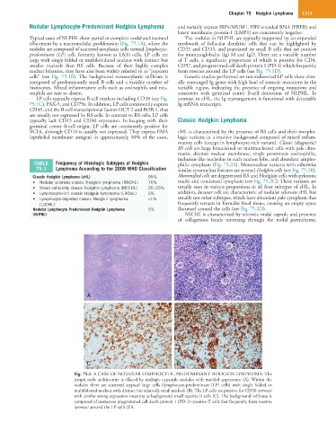
Fig. 75.1 A CASE OF NODULAR LYMPHOCYTE–PREDOMINANT HODGKIN LYMPHOMA. The
lymph node architecture is effaced by multiple expansile nodules with mottled appearance (A). Within the
nodules there are scattered atypical large cells (lymphocyte-predominant [LP] cells) with single folded or
multilobated nucleus with distinct but relatively small nucleoli (B). The LP cells are positive for CD20 (arrows)
with similar strong expression intensity as background small reactive B cells (C). The background infiltrate is
composed of numerous programmed cell death protein 1 (PD-1)−positive T cells that frequently form rosettes
(arrows) around the LP cells (D).