
Page 1449 - Hematology_ Basic Principles and Practice ( PDFDrive )
P. 1449
Chapter 80 Clinical Manifestations, Staging, and Treatment of Follicular Lymphoma 1289
TABLE Incidence Rates Per 100,000 of the Indolent TABLE Mutations in Follicular Lymphoma
80.1 Lymphomas (Ref) 80.2
All Male Female Frequency
Gene Name Abbreviation Location at Diagnosis
Non-hodgkin lymphoma 32.2 40.2 25.9
Myeloid/lymphoid or MLL2 12q 89%
Follicular lymphoma 3.6 3.9 3.3
mixed-lineage leukemia 2
Small B lymphocytic lymphoma/Chronic 6.6 7.9 4.6
lymphocytic leukemia/ CREB binding protein CREBBP 16p 33%
Tumor necrosis factor receptor TNFRSF14 1p 25%
Lymphoplasmacytic lymphoma 0.6 0.8 0.5
superfamily member 14
Mantle cell lymphoma 0.8 1.3 0.4
E1A binding protein p300 EP300 22q 15%
Marginal zone lymphoma 2.0 2.1 1.9
Myocyte enhancer factor 2B MEF2B 19p 13%
From SEER data base https://seer.cancer.gov/csr/1975_2013/browse_csr.php?s
ectionSEL=19&pageSEL=sect_19_table.26.html (accessed January 29, 2017) Enhancer of zeste homolog 2 EZH2 7q 11%
A
BCL6 CD10 BCL2 control BCL2
B C D E
F G H I
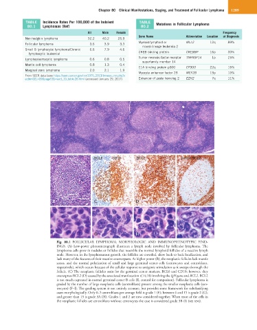
Fig. 80.1 FOLLICULAR LYMPHOMA: MORPHOLOGIC AND IMMUNOPHENOTYPIC FIND-
INGS. (A) Low-power photomicrograph illustrates a lymph node involved by follicular lymphoma. The
lymphoma cells grow in nodules or follicles that resemble the normal lymphoid follicles of a reactive lymph
node. However, in the lymphomatous growth, the follicles are crowded, show back-to-back localization, and
lack many of the features of their reactive counterparts. At higher power (B), the neoplastic follicles lack mantle
zones, and the normal polarization of small and large germinal center cells (centrocytes and centroblasts,
respectively), which occurs because of the cellular response to antigenic stimulation as it sweeps thorough the
follicle. (C) The neoplastic follicles stain for the germinal center markers, BCL6 and CD10; however, they
overexpress BCL2 (D) caused by the associated translocation t(14;18) involving the IgH gene and BCL2. BCL2
is not much expressed in normal germinal center B cells (E, control for comparison). Follicular lymphoma is
graded by the number of large neoplastic cells (centroblasts) present among the smaller neoplastic cells (cen-
trocytes) (F–I). The grading system is not entirely accurate, but provides some framework for subclassifying
cases morphologically. Only 0–5 centroblasts per average field is grade 1 (F); between 6 and 15 is grade 2 (G);
and greater than 15 is grade 3A (H). Grades 1 and 2 are now considered together. When most of the cells in
the neoplastic follicles are centroblasts without centrocytes the case is considered grade 3B (I) (see text).