
Page 1450 - Hematology_ Basic Principles and Practice ( PDFDrive )
P. 1450
1290 Part VII Hematologic Malignancies
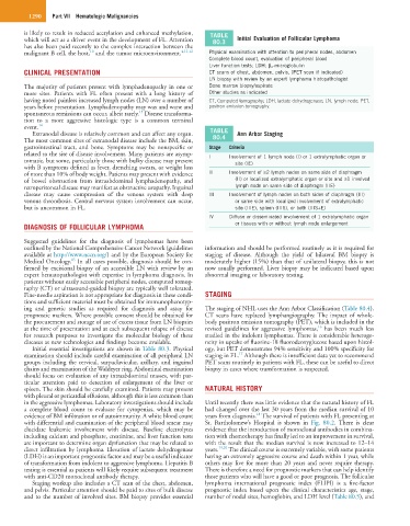
is likely to result in reduced acetylation and enhanced methylation, TABLE
which will act as a driver event in the development of FL. Attention 80.3 Initial Evaluation of Follicular Lymphoma
has also been paid recently to the complex interaction between the
10
malignant B cell, the host, and the tumor microenvironment. 4,11,12 Physical examination with attention to peripheral nodes, abdomen
Complete blood count, evaluation of peripheral blood
Liver function tests; LDH; β 2 -microglobulin
CLINICAL PRESENTATION CT scans of chest, abdomen, pelvis, (PET scan if indicated)
LN biopsy with review by an expert lymphoma histopathologist
The majority of patients present with lymphadenopathy in one or Bone marrow biopsy/aspirate
more sites. Patients with FL often present with a long history of Other studies as indicated
having noted painless increased lymph nodes (LN) over a number of CT, Computed tomography; LDH, lactate dehydrogenase; LN, lymph node; PET,
years before presentation. Lymphadenopathy may wax and wane and positron emission tomography.
13
spontaneous remissions can occur, albeit rarely. Disease transforma-
tion to a more aggressive histologic type is a common terminal
event. 14 TABLE
Extranodal disease is relatively common and can affect any organ. Ann Arbor Staging
The most common sites of extranodal disease include the BM, skin, 80.4
gastrointestinal tract, and bone. Symptoms may be nonspecific or Stage Criteria
related to the site of disease involvement. Many patients are asymp- I Involvement of 1 lymph node (I) or 1 extralymphatic organ or
tomatic, but some, particularly those with bulky disease may present site (IE)
with B symptoms defined as fever, drenching sweats, or weight loss
of more than 10% of body weight. Patients may present with evidence II Involvement of ≥2 lymph nodes on same side of diaphragm
of bowel obstruction from intraabdominal lymphadenopathy, and (II) or localized extralymphatic organ or site and ≥1 involved
retroperitoneal disease may manifest as obstructive uropathy. Inguinal lymph node on same side of diaphragm (IIE)
disease may cause compression of the venous system with deep III Involvement of lymph nodes on both sides of diaphragm (III)
venous thrombosis. Central nervous system involvement can occur, or same side with localized involvement of extralymphatic
but is uncommon in FL. site (IIIE), spleen (IIIS), or both (IIIS+E)
IV Diffuse or disseminated involvement of 1 extralymphatic organ
DIAGNOSIS OF FOLLICULAR LYMPHOMA or tissues with or without lymph node enlargement
Suggested guidelines for the diagnosis of lymphomas have been
outlined by the National Comprehensive Cancer Network (guidelines information and should be performed routinely as it is required for
available at http://www.nccn.org/) and by the European Society for staging of disease. Although the yield of bilateral BM biopsy is
15
Medical Oncology. In all cases possible, diagnosis should be con- moderately higher (15%) than that of unilateral biopsy, this is not
firmed by excisional biopsy of an accessible LN with review by an now usually performed. Liver biopsy may be indicated based upon
expert hematopathologist with expertise in lymphoma diagnosis. In abnormal imaging or laboratory testing.
patients without easily accessible peripheral nodes, computed tomog-
raphy (CT) or ultrasound-guided biopsy are typically well tolerated.
Fine-needle aspiration is not appropriate for diagnosis in these condi- STAGING
tions and sufficient material must be obtained for immunophenotyp-
ing and genetic studies as required for diagnosis and assay for The staging of NHL uses the Ann Arbor Classification (Table 80.4).
prognostic markers. Where possible consent should be obtained for CT scans have replaced lymphangiography. The impact of whole-
the procurement and storage of use of excess tissue from LN biopsies body positron emission tomography (PET), which is included in the
16
at the time of presentation and at each subsequent relapse of disease revised guidelines for aggressive lymphomas, has been much less
for research purposes to investigate the molecular biology of these studied in the indolent lymphomas. There is considerable heteroge-
diseases as new technologies and findings become available. neity in uptake of fluorine-18 fluorodeoxyglucose based upon histol-
Initial essential investigations are shown in Table 80.3. Physical ogy, but PET demonstrates 94% sensitivity and 100% specificity for
17
examination should include careful examination of all peripheral LN staging in FL. Although there is insufficient data yet to recommend
groups including the cervical, surpaclavicular, axillary, and inguinal PET scans routinely in patients with FL, these can be useful to direct
chains and examination of the Waldeyer ring. Abdominal examination biopsy in cases where transformation is suspected.
should focus on evaluation of any intraabdominal masses, with par-
ticular attention paid to detection of enlargement of the liver or
spleen. The skin should be carefully examined. Patients may present NATURAL HISTORY
with pleural or pericardial effusions, although this is less common than
in the aggressive lymphomas. Laboratory investigations should include Until recently there was little evidence that the natural history of FL
a complete blood count to evaluate for cytopenias, which may be had changed over the last 30 years from the median survival of 10
18
evidence of BM infiltration or of autoimmunity. A white blood count years from diagnosis. The survival of patients with FL presenting at
with differential and examination of the peripheral blood smear may St. Bartholomew’s Hospital is shown in Fig. 80.2. There is clear
elucidate leukemic involvement with disease. Baseline electrolytes evidence that the introduction of monoclonal antibodies in combina-
including calcium and phosphate, creatinine, and liver function tests tion with chemotherapy has finally led to an improvement in survival,
are important to determine organ dysfunction that may be related to with the result that the median survival is now increased to 12–14
direct infiltration by lymphoma. Elevation of lactate dehydrogenase years. 19,20 The clinical course is extremely variable, with some patients
(LDH) is an important prognostic factor and may be a useful indicator having an extremely aggressive course and death within 1 year, while
of transformation from indolent to aggressive lymphoma. Hepatitis B others may live for more than 20 years and never require therapy.
testing is essential as patients will likely require subsequent treatment There is therefore a need for prognostic markers that can help identify
with anti-CD20 monoclonal antibody therapy. those patients who will have a good or poor prognosis. The follicular
Staging workup also includes a CT scan of the chest, abdomen, lymphoma international prognostic index (FLIPI) is a five-factor
and pelvis. Particular attention should be paid to sites of bulk disease prognostic index based upon the clinical characteristics age, stage,
and to the number of involved sites. BM biopsy provides essential number of nodal sites, hemoglobin, and LDH level (Table 80.5), and