
Page 1542 - Hematology_ Basic Principles and Practice ( PDFDrive )
P. 1542
Chapter 85 T-Cell Lymphomas 1369
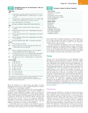
TABLE ISCL/EORTC Revision to the Classification of MF and TABLE Therapeutic Options for Mycosis Fungoides
85.5 SS (Adaptation) 85.6
TNMB Stages Topical Therapy
Skin Ultraviolet A with psoralen
Limited patches, papules, and/or plaques covering <10% of the
T 1 Ultraviolet B
skin surface. May stratify into T 1a (patch only) vs T 1b (plaque External beam radiation therapy
± patch). Total-skin electron beam radiation
Patches, papules or plaques covering ≥10% of skin surface. May Topical chemotherapy
T 2
stratify into T 2a (patch only) vs T 2b (plaque ± patch). Topical retinoids
Systemic Therapy
One or more tumors (≥1-cm diameter)
T 3
Photophoresis
Confluence of erythema covering ≥80% body surface area
T 4 Interferon-α
Node Oral retinoids
No clinically abnormal peripheral lymph nodes; biopsy not
N 0 Targeted therapies
required Single-agent chemotherapy
Abnormal peripheral lymph nodes; histopathology Dutch grade 1 Combination chemotherapy
N 1
Stem cell transplantation
or NCI LN 0–2
Investigational agents
Abnormal peripheral lymph nodes; histopathology Dutch grade 2
N 2
or NCI LN 3
N 3 Abnormal peripheral lymph nodes; histopathology Dutch grades
3–4 or NCI LN 4 from typical small, convoluted lymphocytes to larger lymphocytes,
N x Abnormal peripheral lymph nodes; no histologic confirmation such as those associated with large-cell lymphoma, has been docu-
Visceral mented. Whether this conversion is secondary to prior therapeutic
No visceral organ involvement modalities used remains uncertain.
M 0
The gold standard for the diagnosis of MF is still routine histo-
Visceral involvement (must have pathology confirmation and
M 1 pathologic evaluation with adequate clinical correlation. Early lesions
involved organ should be specified) of MF are frequently accompanied by heavy infiltrates of benign
Blood reactive T cells, hampering the detection of abnormal T-cell clones
Absence of significant blood involvement: ≤5% of peripheral by any laboratory method. Hence most adjuvant laboratory methods
B 0
blood lymphocytes are atypical (Sézary) cells are not helpful at the precise time when they are most needed.
Low blood tumor burden: >5% of peripheral blood lymphocytes
B 1
are atypical (Sézary) but does not meet the criteria for B 2 Therapy
B2 High blood tumor burden: ≥1000/µL Sézary cells with positive
clones Therapy can be conveniently divided into two approaches: topical
Clinical Staging (skin directed), such as psoralen plus ultraviolet A (PUVA), topical
IA T1, N0, M0, B0,1 chemotherapy application (nitrogen mustard or carmustine), external
IB T2, N0, M0, B0,1 beam radiotherapy, and total-skin electron beam radiotherapy, and
IIA T1,2, N1,2, M0, B0,1 systemic (skin and viscera directed), such as IFNs, oral or parenteral
IIB T3, N0–2, M0, B0,1 chemotherapy, photopheresis, oral retinoids, and investigational new
IIIA T4, N0–2, M0, B0 compounds (Table 85.6). No studies have demonstrated that one
IIIB T4, N0–2, M0, B1 topical therapy is more effective than another, and patient and
investigator preference remains the most important discriminating
T1–4, N0–2, M0, B2
IVA 1 factor governing choice. However, as the biology of the neoplastic
T1–4, N3, M0, B0–2
IVA 2 cell has become better understood, it is clear that some therapies may
IVB T1–4, N0–3, M1, B0–2 actually have topical and systemic effects through alterations in the
CTCL, Cutaneous T-cell lymphoma; EORTC, European Organization of Research body’s cytokine milieu and ability to mount a host response against
and Treatment of Cancer; ISCL, International Society for Cutaneous the neoplastic cell. Furthermore, knowledge regarding signaling
Lymphomas; NCI LN, National Cancer Institute lymph node grading system; pathways in the neoplastic cells in MF/SS offers hope that future
TNMB, tumor-node-metastasis-blood.
development of targeted treatment approaches will soon be available.
Investigational approaches combining therapies also remains an active
research strategy (see box on How We Manage Mycosis Fungoides/
allows the calculation of a value based on the degree of nuclear Sézary Syndrome).
folding; this nuclear contour index is significantly greater in patients
with MF than in other benign conditions.
The density of epidermal Langerhans cells in biopsy samples, as Phototherapy
determined by immunoperoxidase stains, has been identified as a
prognostic feature. Epidermal Langerhans cells are necessary for 8-Methoxypsoralen (8-MOP) is a member of a family of photoacti-
antigen recognition and processing in the normal immune response. vated compounds (furocoumarin derivatives), which may inhibit
2
Patients with Langerhans cell densities greater than 90 cells/mm had DNA and RNA synthesis through formation of monofunctional or
a significantly reduced risk for death from MF/SS compared with bifunctional thymine adducts, gene mutations, or sister chromatid
those with lower densities. There was no prognostic significance exchanges. The cross-strand formed between DNA strands results in
+
identified for the presence or absence of CD30 cells. It has been a halt in cell division, as well as oxidative damage to cytoplasmic
+
noted that the presence of cytotoxic CD8 T lymphocytes in the skin organelles and cell membranes. These drugs are active only if the
infiltrate is associated with a more favorable prognosis. tissue containing the psoralen compound is exposed to ultraviolet A
Occasionally patients may develop a more clinically aggressive (UVA). The mechanism of cell cytotoxicity for many cancer therapies
lymphoma concurrent with a change in the histologic appearance of involves the induction of apoptosis. Yoo et al demonstrated that
the neoplastic cells and the pace of their disease. This progression peripheral blood mononuclear cells from SS patients and controls