
Page 1539 - Hematology_ Basic Principles and Practice ( PDFDrive )
P. 1539
1366 Part VII Hematologic Malignancies
Differential Diagnosis presentation. These primary cutaneous CD30 LCLs are probably
+
closely related to LyP, regressing atypical histiocytosis, and primary
Primary CTCL represents a heterogeneous group of disorders with cutaneous HL. The tumor has a favorable prognosis, and often
considerable variability in histologic characteristics, phenotype, and complete or partial spontaneous regression occurs. This is in contrast
+
prognosis. The Kiel Classification, the Working Formulation, and the to primary noncutaneous CD30 LCLs, which can be seen in children
Revised European-American Lymphoma (REAL) classification system or adults and which carries a poor prognosis. These primary cutane-
were developed for NHLs and were not designed to provide an ous lesions, in contrast to nodal or pediatric cases, have been shown
adequate characterization of the spectrum of CTCLs. To address the to rarely have the chromosomal translocation t(2;5) associated with
deficiencies of the previously proposed systems, a more clinically overexpression of ALK (ALK negative). Histopathology consists of
useful classification was developed by the European Organization for diffuse nonepidermotropic infiltrates with cohesive sheets of large
+
Research and Treatment of Cancer (EORTC). WHO has proposed CD30 tumor cells (Fig. 85.20). In most instances, the tumor cells
a classification with nearly 90% concordance with the EORTC clas- have anaplastic morphologic characteristics, showing round, oval, or
sification (see Table 85.4). A number of other disorders in which irregularly shaped nuclei; prominent (eosinophilic) nucleoli; and
malignant T cells infiltrate the skin should be distinguished from abundant cytoplasm. Less commonly, the neoplastic cells have a
MF/SS. These disorders are discussed in the following sections. pleomorphic or immunoblastic appearance. Reactive lymphocytes are
often present, but infiltrating eosinophils are often less conspicuous.
+
The immunophenotype of this disorder is characteristically CD4 ,
CD30 Lymphoproliferative Disorders with more than 75% of neoplastic cells expressing CD30. In contrast
+
to the poor outcome of MF that has transformed to a CD30 large-
+
CD30 lymphoproliferative disorders include LyP, primary cutaneous cell variant, primary cutaneous CD30 LCLs are associated with an
ALCL, and a spectrum of borderline cases. By definition, ALCL excellent prognosis. Radiotherapy is the preferred treatment for soli-
presents with single or multiple tumors measuring more than 2 cm tary or localized disease, with combination chemotherapy reserved
and with a tendency for ulceration and steady growth. Borderline for patients with generalized skin lesions or extracutaneous dissemi-
lesions are smaller but also tend to have a prolonged course, often nation. Surgical excision may be adequate in many cases. In advanced
with spontaneous resolution. cases, 5-year survival exceeds 30%.
−
Lymphomatoid Papulosis CD30 Cutaneous T-Cell Lymphoma
LyP is characterized by recurrent crops of self-healing, red-brown, These rare presentations often classified as CTCL, NOS, or d’emblée
centrally necrotic, asymptomatic papules and nodules. This entity presentation, tend to have an aggressive clinical course. Patients
represents 10%–15% of all CTCL cases. Patients may have a few present with localized or generalized plaques, nodules, or tumors.
lesions or more than 100 at a time. Histologic evaluation reveals an
+
atypical CD4 lymphocytic infiltrate with a variable mixed inflam-
matory infiltrate. These may be primarily small cerebriform cells
similar to those seen in MF (type B), but most often there are larger
+
CD30 cells with prominent nucleoli resembling Reed-Sternberg cells
(type A). A third variety of LyP (type C), also considered borderline
+
ALCL, presents with sheets of large cells resembling CD30 large
cells. TCR gene rearrangement studies demonstrate a clonal origin.
Although the typical course is usually indolent, spanning decades,
approximately 15% of patients develop MF, cutaneous ALCL, and
very rarely HL or NHL during their lifetime. A direct link between
LyP, CTCL, and HL was demonstrated in a patient with the three
lymphoproliferative disorders arising from a common T-cell clone, as
shown by TCR gene studies.
CD30 Cutaneous T-Cell Lymphoma
+
+
Primary cutaneous CD30 LCL typically occurs in adults presenting
+
with solitary or localized (ulcerating) nodules or tumors (Fig. 85.19). Fig. 85.19 LESIONS OF CD30 LARGE-CELL LYMPHOMA WITH
Regional lymph node involvement is seen in 25% of patients at ULCERATION.
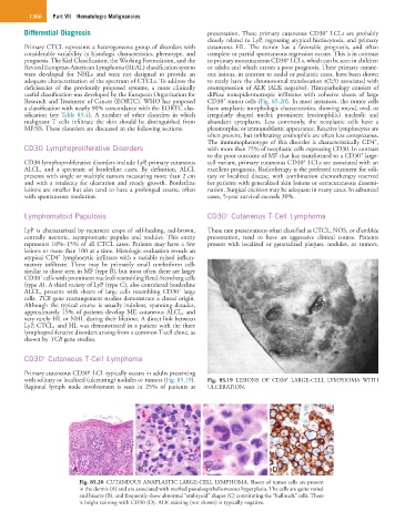
A B C D
Fig. 85.20 CUTANEOUS ANAPLASTIC LARGE-CELL LYMPHOMA. Sheets of tumor cells are present
in the dermis (A) and are associated with marked pseudoepitheliomatous hyperplasia. The cells are quite varied
and bizarre (B), and frequently show abnormal “embryoid” shapes (C) constituting the “hallmark” cells. There
is bright staining with CD30 (D). ALK staining (not shown) is typically negative.